. 2017 Jun 5;8(1):131.
doi: 10.1186/s13287-017-0575-5.

Human liver mesenchymal stem/progenitor cells inhibit hepatic stellate cell activation: in vitro and in vivo evaluation

Affiliations

Human liver mesenchymal stem/progenitor cells inhibit hepatic stellate cell activation: in vitro and in vivo evaluation

Mustapha Najimi et al. Stem Cell Res Ther. .

Abstract

Background: Progressive liver fibrosis leads to cirrhosis and end-stage liver disease. This disease is a consequence of strong interactions between matrix-producing hepatic stellate cells (HSCs) and resident and infiltrating immune cell populations. Accumulated experimental evidence supports the involvement of adult-derived human liver mesenchymal stem/progenitor cells (ADHLSCs) in liver regeneration. The aim of the present study was to evaluate the influence of ADHLSCs on HSCs, both in vitro and in vivo.

Methods: Activated human HSCs were co-cultured with ADHLSCs or ADHLSC-conditioned culture medium. The characteristics of the activated human HSCs were assessed by microscopy and biochemical assays, whereas proliferation was analyzed using flow cytometry and immunocytochemistry. The secretion profile of activated HSCs was evaluated by ELISA and Luminex. ADHLSCs were transplanted into a juvenile rat model of fibrosis established after co-administration of phenobarbital and CCl4.

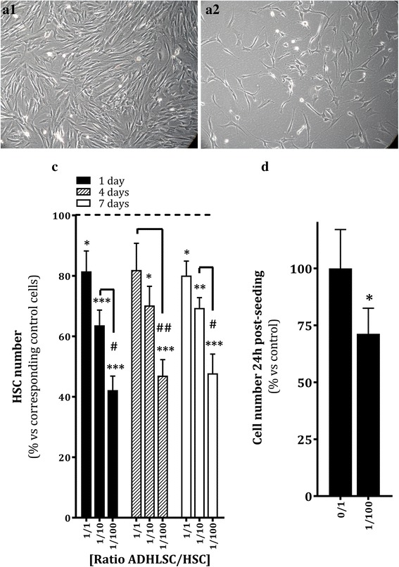
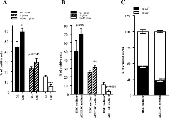
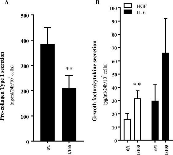
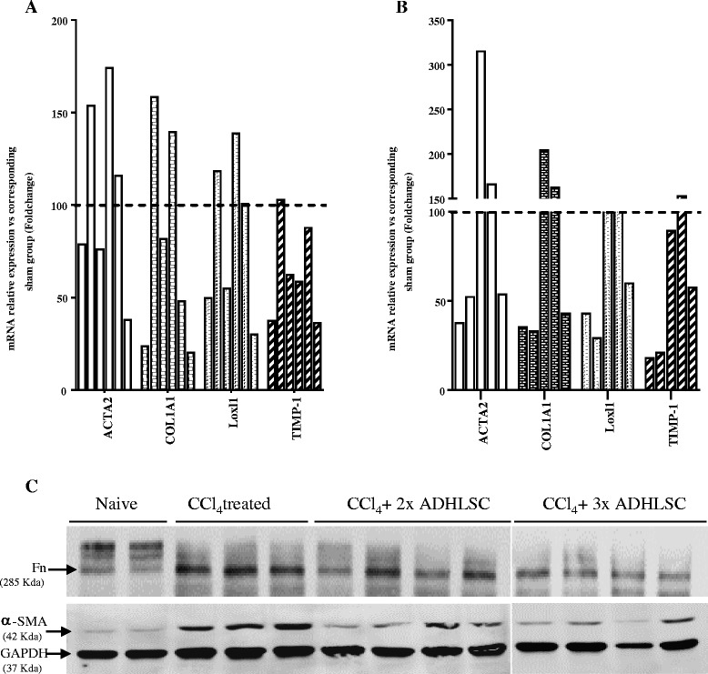
Results: When co-cultured with ADHLSCs or conditioned medium, the proliferation of HSCs was inhibited, beginning at 24 h and for up to 7 days. The HSCs were blocked in G0/G1 phase, and showed decreased Ki-67 positivity. Pro-collagen I production was reduced, while secretion of HGF, IL-6, MMP1, and MMP2 was enhanced. Neutralization of HGF partially blocked the inhibitory effect of ADHLSCs on the proliferation and secretion profile of HSCs. Repeated intrahepatic transplantation of cryopreserved/thawed ADHLSCs without immunosuppression inhibited the expression of markers of liver fibrosis in 6 out of 11 rats, as compared to their expression in the vehicle-transplanted group.

Conclusions: These data provide evidence for a direct inhibitory effect of ADHLSCs on activated HSCs, which supports their development for the treatment of liver fibrosis.

Keywords: Hepatic stellate cells; Liver; Liver fibrosis; Liver stem/progenitor cells; Secretome.


Figures

Fig. 1
Fig. 1
Effect of ADHLSCs on HSC culture. a Morphology of HSCs recovered after 24 h of co-culture without ADHLSCs (A1) or with ADHLSCs at an ADHLSC:HSC ratio of 1:100 (A2). b Significant decrease in the number of adherent HSCs after 24 h of co-culture at an ADHLSC:HSC ratio of 1:100, which was maintained for up to 7 days (N = 6 for 24 h; N = 4 for 4 and 7 days). c Significant decrease in the number of HSCs after 24 h of co-culture at an ADHLSC:HSC ratio of 1:100, using the CCK-8 biochemical assay (N = 4). Results are expressed as mean ± standard error of the mean (SEM). *** p value < 0.001; ** p < 0.01; * p < 0.05. ADHLSCs adult-derived human liver mesenchymal stem/progenitor cells, HSC hepatic stellate cells
Fig. 2
Fig. 2
Effect of ADHLSCs on HSC proliferation. Cell cycle analysis of PI-stained HSCs demonstrated an increase in the number of HSC blocked in G0/G1 phase and a decrease in the number of HSCs in G2/M phase after 24 h of co-culture at an ADHLSC:HSC ratio of 1:100 (a) and after 24 h of incubation with ADHLSC-conditioned medium (b) (N = 4). c The Ki67 immunostaining of HSCs showed a significant decrease in the number of immunostained nuclei of HSCs pre-incubated for 24 h with ADHLSC conditioned medium (N = 4). For each experimental condition, four different fields were analyzed, and a total of 2500 nuclei were counted using Image J software. Results are expressed as mean ± standard error of the mean (SEM). *** p value < 0.001; ** p < 0.01; * p < 0.05. ADHLSCs adult-derived human liver mesenchymal stem/progenitor cells, HSC hepatic stellate cells
Fig. 3
Fig. 3
Modulation of the HSC secretome after 24 h of co-culture with ADHLSCs. a ADHLSCs inhibit pro-collagen type I secretion by HSCs after 24 h of co-culture at a ADHLSC: HSC ratio of 1:100, as evaluated by ELISA (N = 4). b The presence of ADHLSCs increased HGF and IL-6 secretion by HSCs after 24 h of co-culture at an ADHLSC:HSC ratio of 1:100, as evaluated by multiplex technology (N = 4). Results are expressed as mean ± standard error of the mean (SEM). *** p value < 0.001; ** p < 0.01; * p < 005. HGF hepatocyte growth factor, IL-6 interleukin 6
Fig. 4
Fig. 4
Involvement of HGF in the effect of ADHLSCs on HSC proliferation and procollagen type I secretion. a The numbers of floating and adhering HSCs after 24 h of incubation with ADHLSC- or HSC-conditioned medium was evaluated after pre-incubation with or without an anti-HGF antibody. There was significant, partial inhibition of the decrease in the number of adhering HSCs when incubated with ADHLSC-conditioned medium containing anti-HGF antibody (N > 5). b Inhibition of the effect of ADHLSC-conditioned medium on HSC pro-collagen type I secretion in the presence of an anti-HGF antibody (N = 3). c Anti-HGF antibody significantly inhibited the increase in HGF, IL-6, MMP1, and MMP2 secretion by HSCs. HGF hepatocyte growth factor, HSC hepatic stellate cells
Fig. 5
Fig. 5
Effect of ADHLSC transplantation on the expression of HSC activation markers in an established model of liver fibrosis. The mRNA levels of αSMA, Col1α1, Lox, and TIMP-1 in the livers of rats intrahepatically transplanted twice (n = 6) (a) or three times (n = 5) (b) with ADHLSCs without immunosuppression were estimated using RT-qPCR. c Total protein extracts were prepared from the three groups of rats as well as from naïve controls (n = 2), and immunoblotted with antibodies against markers of HSC activation and GAPDH as described in the “Materials and Methods” section. Downregulation of α-SMA, and fibronectin (Fn) is correlated to ADHLSC transplantation twice (n = 4) and three times (n = 4). ADHLSCs adult-derived human liver mesenchymal stem/progenitor cells

Similar articles

Cited by

References

    1. Friedman SL. Liver fibrosis -- from bench to bedside. J Hepatol. 2003;38(Suppl 1):S38–53. doi: 10.1016/S0168-8278(02)00429-4. - DOI - PubMed
    1. Friedman SL. Hepatic fibrosis - overview. Toxicology. 2008;254(3):120–9. doi: 10.1016/j.tox.2008.06.013. - DOI - PubMed
    1. Francoz C, Belghiti J, Durand F. Indications of liver transplantation in patients with complications of cirrhosis. Best Pract Res Clin Gastroenterol. 2007;21(1):175–90. doi: 10.1016/j.bpg.2006.07.007. - DOI - PubMed
    1. Evans HM, Kelly DA, McKiernan PJ, Hubscher S. Progressive histological damage in liver allografts following pediatric liver transplantation. Hepatology. 2006;43(5):1109–17. doi: 10.1002/hep.21152. - DOI - PubMed
    1. Li JT, Liao ZX, Ping J, Xu D, Wang H. Molecular mechanism of hepatic stellate cell activation and anti-fibrotic therapeutic strategies. J Gastroenterol. 2008;43(6):419–28. doi: 10.1007/s00535-008-2180-y. - DOI - PubMed

MeSH terms