Review
. 2017 Oct:139:115-122.
doi: 10.1016/j.visres.2017.04.014. Epub 2017 Jun 28.

Cell-cell communication in diabetic retinopathy

Affiliations
Review

Cell-cell communication in diabetic retinopathy

Sayon Roy et al. Vision Res. 2017 Oct.

Abstract

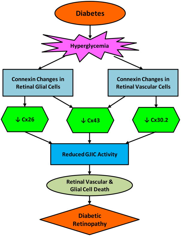
In diabetic retinopathy, high glucose (HG)-mediated breakdown in cell-cell communication promotes disruption of retinal homeostasis. Several studies indicate that HG condition alters expression of connexin genes and subsequent gap junction intercellular communication (GJIC) in retinal vascular cells and non-vascular cells. A serious consequence of disrupted cell-cell communication is apoptosis and breakdown of the blood-retinal barrier (BRB). More recently, studies suggest adverse effects from HG on retinal Müller cells. This article focuses on HG-mediated changes in connexin expression and GJIC and their subsequent effects on the breakdown of retinal homeostasis, cell death, compromised vascular permeability, and interactions between endothelial cells, pericytes and retinal Müller cells in the pathogenesis of diabetic retinopathy. Additionally, options for rectifying disrupted homeostasis under HG condition associated with diabetic retinopathy are reviewed.

Keywords: Connexins; Diabetic retinopathy; Gap junctions.


Figures

Figure 1
Figure 1. Structure and localization of connexons
Each connexon is a hexameric channel comprised of six homomeric or heteromeric subunits. Connexons are localized on the cell surface; each connexon exhibits a cytoplasmic domain, a transmembrane domain, and an extracellular domain. The extracellular domain from two connexons play a critical role in docking and represent the “gap” junction or the space between the two cells where connexons form a connexin channel.
Figure 2
Figure 2. Docking of connexons leads to coupled junctions
Gap junction intercellular communication is dependent on successful docking of two connexons located on two adjacent cells. When connexons present on the cell surface of one cell dock with connexons present on the cell surface of an adjacent cell it can lead to interlocking of the extracellular domains of each connexon allowing the formation of a gap junction connexin channel between two adjacent cells. These channels allow exchange of small molecules and ions permitting cell to cell communication.
Figure 3
Figure 3. High glucose (HG)-induced connexin changes in the retinal vascular and glial cells
Retinal glial cells undergo Cx26 and Cx43 downregulation whereas retinal vascular cells undergo Cx43 and Cx30.2 downregulation. HG-induced connexin downregulation in the retina leads to a decrease in GJIC activity, promoting cell loss, ultimately contributing to vision loss in diabetic retinopathy.

References

    1. Altevogt BM, Paul DL. Four classes of intercellular channels between glial cells in the CNS. J Neurosci. 2004;24(18):4313–4323. - PMC - PubMed
    1. Bloomfield SA, Volgyi B. The diverse functional roles and regulation of neuronal gap junctions in the retina. Nat Rev Neurosci. 2009;10(7):495–506. - PMC - PubMed
    1. Chistiakov DA. Diabetic retinopathy: pathogenic mechanisms and current treatments. Diabetes Metab Syndr. 2011;5(3):165–172. - PubMed
    1. Cottrell GT, Burt JM. Functional consequences of heterogeneous gap junction channel formation and its influence in health and disease. Biochim Biophys Acta. 2005;1711(2):126–141. - PubMed
    1. Cui H, Liu Y, Qin L, Wang L, Huang Y. Increased membrane localization of pannexin1 in human corneal synaptosomes causes enhanced stimulated ATP release in chronic diabetes mellitus. Medicine (Baltimore) 2016;95(49):e5084. - PMC - PubMed