Review
. 2017 Nov 10;429(22):3500-3524.
doi: 10.1016/j.jmb.2017.05.027. Epub 2017 Jun 3.

Proteasome Structure and Assembly

Affiliations
Review

Proteasome Structure and Assembly

Lauren Budenholzer et al. J Mol Biol. .

Abstract

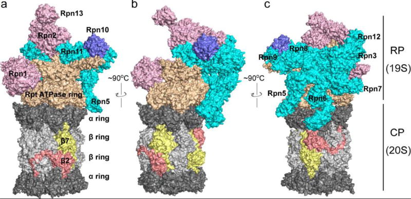
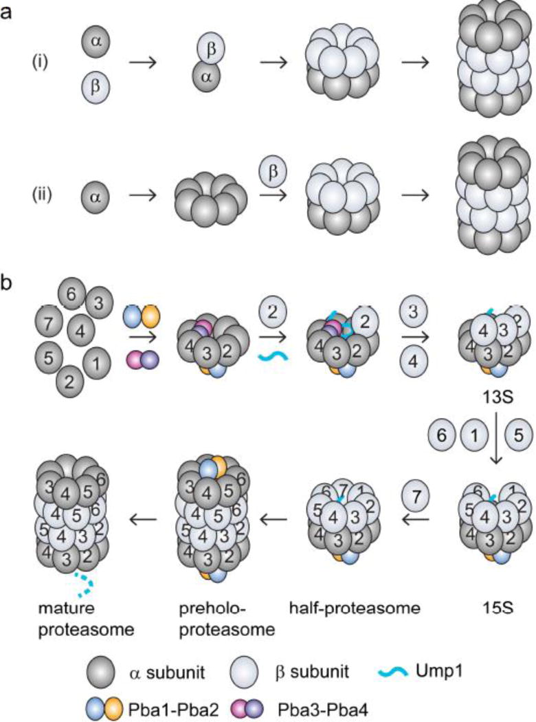
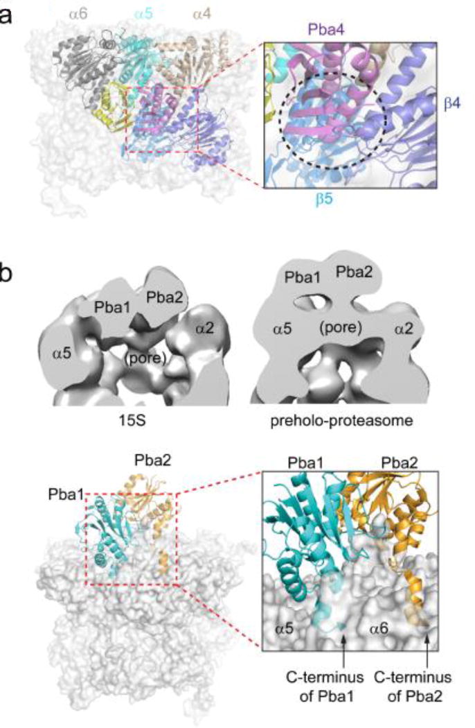
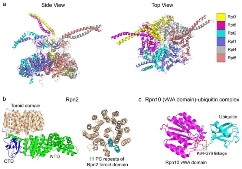
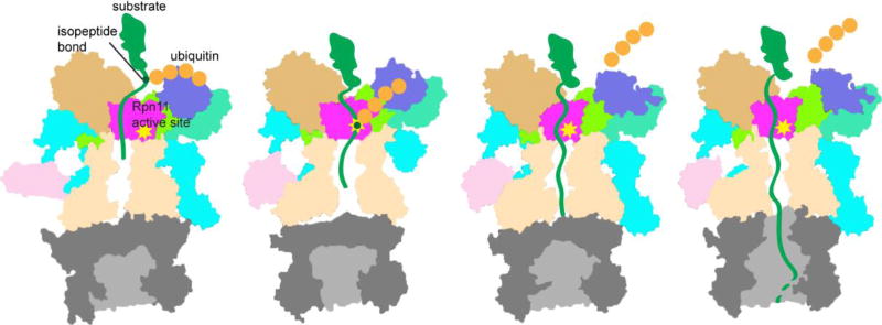
The eukaryotic 26S proteasome is a large multisubunit complex that degrades the majority of proteins in the cell under normal conditions. The 26S proteasome can be divided into two subcomplexes: the 19S regulatory particle and the 20S core particle. Most substrates are first covalently modified by ubiquitin, which then directs them to the proteasome. The function of the regulatory particle is to recognize, unfold, deubiquitylate, and translocate substrates into the core particle, which contains the proteolytic sites of the proteasome. Given the abundance and subunit complexity of the proteasome, the assembly of this ~2.5MDa complex must be carefully orchestrated to ensure its correct formation. In recent years, significant progress has been made in the understanding of proteasome assembly, structure, and function. Technical advances in cryo-electron microscopy have resulted in a series of atomic cryo-electron microscopy structures of both human and yeast 26S proteasomes. These structures have illuminated new intricacies and dynamics of the proteasome. In this review, we focus on the mechanisms of proteasome assembly, particularly in light of recent structural information.

Keywords: proteasome; proteasome assembly; protein degradation; ubiquitin proteasome system.


Figures

Figure 1
Figure 1
The structure of the 26S proteasome. The CP is in gray with α rings in dark gray and β rings in light gray. The Rpt ATPase ring subunits of the RP base are colored light brown, while the Rpn1, Rpn2 and Rpn13 base subunits are pink. Individual lid subunits labeled and depicted in cyan, and Rpn10 is blue. The CP subunits β2 and β7 are depicted in copper and yellow, respectively, to highlight their C-terminal tails that extend to neighboring subunits. The positions of individual CP and base subunits are indicated in Figure 2 and Figure 4 due to space limitations in Figure 1. Figures were generated from PDB 3JCP.
Figure 2
Figure 2
Models for CP assembly pathways. (a) Two proposed assembly pathways are the (i) α-ring-independent (or α-β heterodimer-initiated) pathway and (ii) α-ring-dependent pathway. (b) Assembly pathway of the constitutive CP in eukaryotes. The α-ring is assembled with the help of two pairs of chaperone proteins: Pba3–Pba4 and Pba1–Pba2. β2 and Ump1 then associate with the α-ring. The association of β3 and β4 is accompanied by the dissociation of Pba3–Pba4. Then β5, β6 and β1 are incorporated sequentially, followed by β7. The insertion of β7 leads to dimerization of two half-proteasomes to form the preholoproteasome. In this complex, Pba1–Pba2 and Ump1 are still associated. In the last step, Pba1–Pba2 dissociates from the CP, the propeptides of the β subunits undergo autoprocessing to activate the CP, and Ump1 is degraded, forming the mature CP.
Figure 3
Figure 3
Two pairs of CP assembly chaperone proteins. (a) Pba3–Pba4 associates with α5 and makes contacts with α4 and α6. The steric clash of Pba3–Pba4 with β4 and β5 is shown. Yeast Pba3–Pba4-α5 X-ray structure was modeled into the full CP structure by superimposing α5 from the two structures. The view is from the axial channel of the CP. α4: light brown; α5: cyan; α6: in dark gray; β4: purple; β5: light blue; Pba3: yellow; Pba4: magenta. The black circle in the inset highlights the steric conflict of Pba3–Pba4 with β4 and β5. Figures were generated from Pba3–Pba4-α5 (PDB ID: 2Z5C) and yeast CP (PDB ID: 5CZ4) structures. (b) Different positions of Pba1–Pba2 during CP assembly. Top left: Negative-stain EM structure of the 15S intermediate. Pba1–Pba2 locates in the widened pore of the α-ring. The figure was generated from EMD-2656 using Chimera. Top right: Negative-stain EM structure of the preholoproteasome. Pba1–Pba2 sits on top of the α-ring. The figure was generated using EMD-2658. Bottom left: X-ray crystal structure of a reconstituted Pba1–Pba2-20S complex. Bottom right: highlight of the insertion of the HbYX motifs of Pba1 and Pba2 into CP α-ring pockets. The C-termini of Pba1 and Pba2 are marked with arrows (PDB ID: 4G4S).
Figure 4
Figure 4
Structures of the Rpt heterohexameric ring, Rpn2 and an Rpn10-ubiquitin conjugate. (a) Top and side views of the S. cerevisiae Rpt hexameric ring. The side view reveals that the ring arranges into two roughly co-axial rings or spirals. The upper ring of OB-folds follows the coiled-coils, extending upward, that join pairs of ATPases. The lower tier consists of the AAA-ATPase and C-terminal domains. The top view shows the narrow central pore of the Rpt ring through which substrates are threaded (PDB ID: 5MP9). (b) Crystal structure of yeast Rpn2. The structure on the left illustrates the N-terminal, C-terminal and toroidal domains of Rpn2. The structure on the right highlights a single repeat (in cyan) of the eleven PC repeats of the toroidal domain (PDB ID: 4ADY). (c) Crystal structure of yeast Rpn10 vWA domain in complex with ubiquitin. The isopeptide bond formed between Ubiquitin-G76 and Rpn10-K84 residues is boxed (PDB ID: 5LN1).
Figure 5
Figure 5
Crystal structures of base assembly chaperones alone or in complex with their respective Rpt partners. All chaperones and Rpt subunits depicted are from S. cerevisiae with the exception of the Rpt5ATPase-CTD. The Rpt5ATPase-CTD construct consists of the ATPase domain from Pyrococcus furiosus PAN (an archaeal homolog of Rpt subunits) followed by the C-terminal peptide from S. cerevisiae Rpt5. Regions highlighted in red on Nas6, Rpn14, and Hsm3 represent a single structural repeat of the chaperones. PDB IDs: 2DZN (Nas6-Rpt3CTD), 3WHL (Nas2NTD-Rpt5ATPase-CTD), 3ACP (Rpn14) and 4A3V (Hsm3-Rpt1CTD).
Figure 6
Figure 6
Models of proteasome base assembly in (a) yeast and (b) human. The models are based primarily on characterization of complexes isolated from cells and analyzed by mass spectrometry. The ordering of events is therefore speculative and distinct assembly pathways remain possible. Rpt1–Rpt6 subunits are numbered 1–6 in the figure. Potential differences can be seen in the association order of Rpt dimer complexes in S. cerevisiae and human, but both pathways may occur in both species.
Figure 7
Figure 7
Hierarchical assembly pathway model for the RP lid. In the model, the lid assembles along a two-pronged pathway in which the subcomplexes LP3 and Module 1 are formed first, and then associate to form LP2. The final step in assembly occurs when Rpn12 joins LP2 to form the mature lid. The full lid, but not LP2, is able to bind the free base. PCI subunits (gray) are labeled with the yeast Rpn subunit numbers.
Figure 8
Figure 8
Structure of the recombinant RP lid. (a) The high-resolution cryoEM structure of free lid (PDB ID 3JCK). As in Fig. 7, Rpn8 is shown in purple, Rpn11 in pink, and Sem1 in gold. The six PCI proteins are shown in cyan. (b) The Rpn8–Rpn11 heterodimer (PDB ID 4O8X). Rpn11, light grey, which harbors the DUB activity of the lid, is shown in the context of the Rpn8–11 heterodimer. Active site residues are shown in light pink, and the Zn2+ in yellow. The zinc atom is not depicted to scale for clarity. The Rpn11 active site is easily accessible. (c) The active site of Rpn11 is depicted in the context of the full lid. The inset shows the interactions among Rpn11 (pink), Rpn8 (purple), and Rpn5 (cyan), blocking the Rpn11 active site. Again, the zinc atom is not depicted to scale for clarity.
Figure 9
Figure 9
Model of substrate degradation by the proteasome based on cryo-EM structures of the human proteasome[167]. Four conformations of the proteasome, named SA – SD, are depicted. Figures were made based on PDB entries 5T0G, 5T0H, 5T0I and 5T0J using Pymol to show the cutaway surface of each state.

References

    1. Saeki Y, Tanaka K. Assembly and Function of the Proteasome. Ubiquitin Family Modifiers and the Proteasome. 2012;832:315–337. doi: 10.1007/978-1-61779-474-2_22. - DOI - PubMed
    1. Hochstrasser M. Ubiquitin-dependent protein degradation. Annu. Rev. Genet. 1996;30:405–439. doi: 10.1146/annurev.genet.30.1.405. - DOI - PubMed
    1. Tomko RJ, Jr, Hochstrasser M. Molecular Architecture and Assembly of the Eukaryotic Proteasome. Annu. Rev. Biochem. 2013;82:415–445. doi: 10.1146/annurev-biochem-060410-150257. - DOI - PMC - PubMed
    1. Wehmer M, Sakata E. Recent advances in the structural biology of the 26S proteasome. Int. J. Biochem. Cell Biol. 2016;79:437–442. doi: 10.1016/j.biocel.2016.08.008. - DOI - PubMed
    1. Lu Y, Lee B-H, King RW, Finley D, Kirschner MW. Substrate degradation by the proteasome: A single-molecule kinetic analysis. Science. 2015;348:1250834–1250834. doi: 10.1126/science.1250834. - DOI - PMC - PubMed

Publication types

MeSH terms

Substances

LinkOut - more resources