

Etiology of epithelial barrier dysfunction in patients with type 2 inflammatory diseases
- PMID: 28583447
- PMCID: PMC5753806
- DOI: 10.1016/j.jaci.2017.04.010
Etiology of epithelial barrier dysfunction in patients with type 2 inflammatory diseases
Abstract
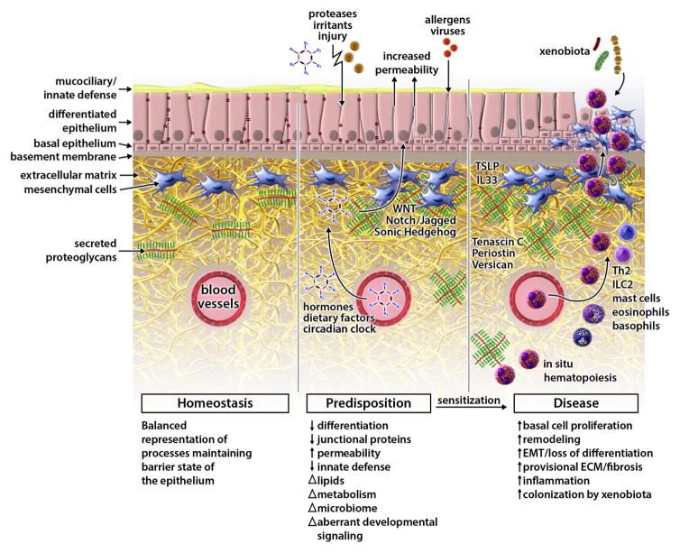
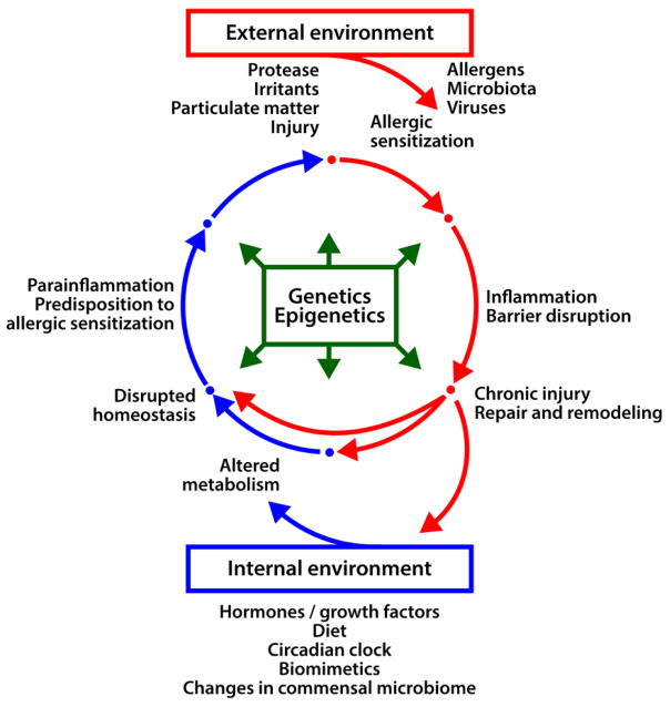
Epithelial barriers of the skin, gastrointestinal tract, and airway serve common critical functions, such as maintaining a physical barrier against environmental insults and allergens and providing a tissue interface balancing the communication between the internal and external environments. We now understand that in patients with allergic disease, regardless of tissue location, the homeostatic balance of the epithelial barrier is skewed toward loss of differentiation, reduced junctional integrity, and impaired innate defense. Importantly, epithelial dysfunction characterized by these traits appears to pre-date atopy and development of allergic disease. Despite our growing appreciation of the centrality of barrier dysfunction in initiation of allergic disease, many important questions remain to be answered regarding mechanisms disrupting normal barrier function. Although our external environment (proteases, allergens, and injury) is classically thought of as a principal contributor to barrier disruption associated with allergic sensitization, there is a need to better understand contributions of the internal environment (hormones, diet, and circadian clock). Systemic drivers of disease, such as alterations of the endocrine system, metabolism, and aberrant control of developmental signaling, are emerging as new players in driving epithelial dysfunction and allergic predisposition at various barrier sites. Identifying such central mediators of epithelial dysfunction using both systems biology tools and causality-driven laboratory experimentation will be essential in building new strategic interventions to prevent or reverse the process of barrier loss in allergic patients.
Keywords: Epithelial barrier; allergic predisposition; allergic sensitization; differentiation; extracellular matrix; hormones; mesenchyme; metabolism; tight junctions.
Copyright © 2017 The Authors. Published by Elsevier Inc. All rights reserved.
Figures
References
-
- Thyssen JP, Kezic S. Causes of epidermal filaggrin reduction and their role in the pathogenesis of atopic dermatitis. J Allergy Clin Immunol. 2014;134:792–9. - PubMed
-
- Pellerin L, Henry J, Hsu CY, Balica S, Jean-Decoster C, Mechin MC, et al. Defects of filaggrin-like proteins in both lesional and nonlesional atopic skin. J Allergy Clin Immunol. 2013;131:1094–102. - PubMed
-
- Palmer CN, Irvine AD, Terron-Kwiatkowski A, Zhao Y, Liao H, Lee SP, et al. Common loss-of-function variants of the epidermal barrier protein filaggrin are a major predisposing factor for atopic dermatitis. Nat Genet. 2006;38:441–6. - PubMed
Publication types
MeSH terms
Grants and funding
LinkOut - more resources
Full Text Sources
Other Literature Sources
Medical
