. 2017 Jul 28;292(30):12471-12482.
doi: 10.1074/jbc.M116.763565. Epub 2017 Jun 5.

Allosteric modulation of human dopamine transporter activity under conditions promoting its dimerization

Affiliations

Allosteric modulation of human dopamine transporter activity under conditions promoting its dimerization

Mary Hongying Cheng et al. J Biol Chem. .

Abstract

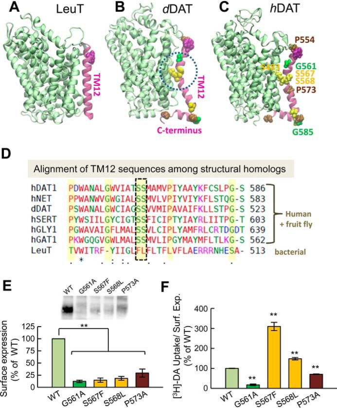
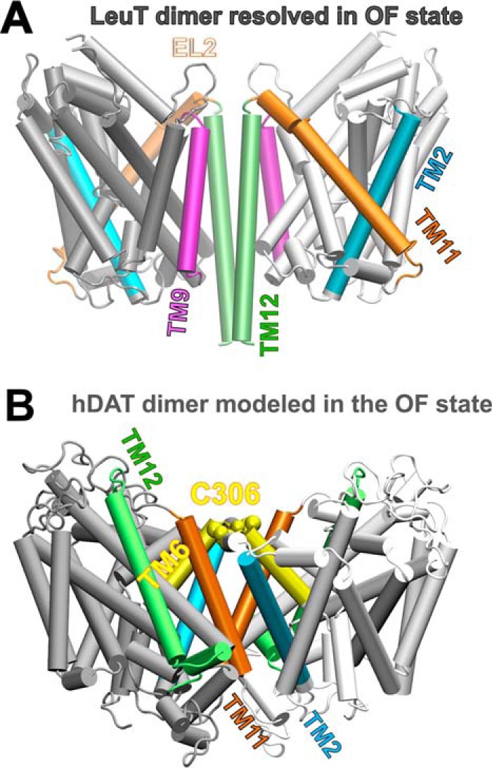
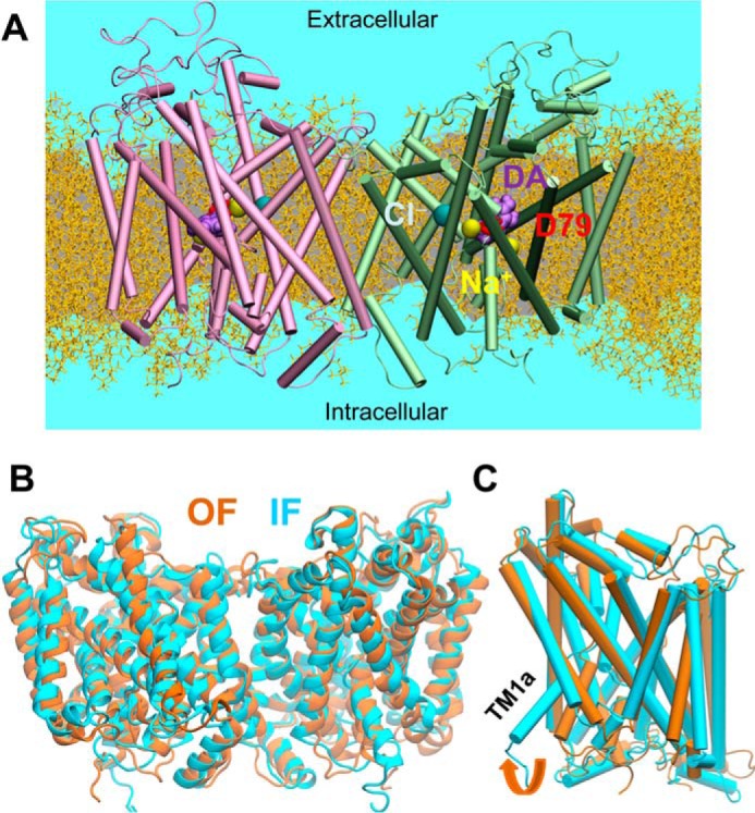
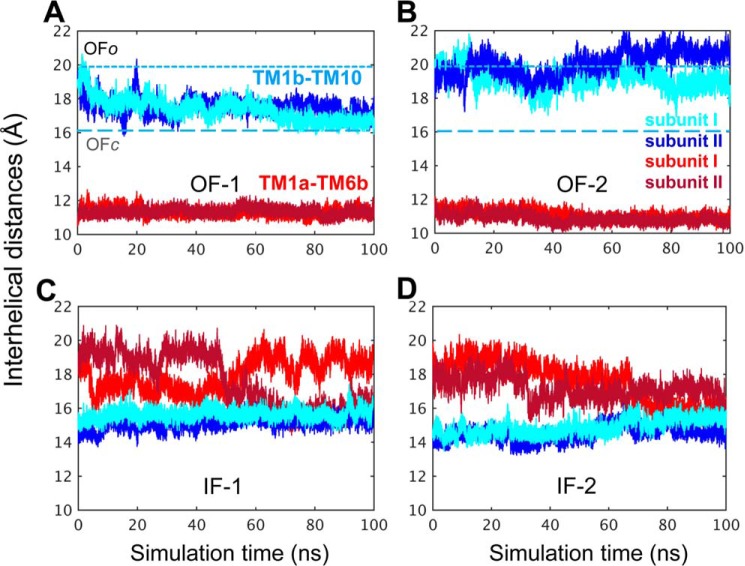
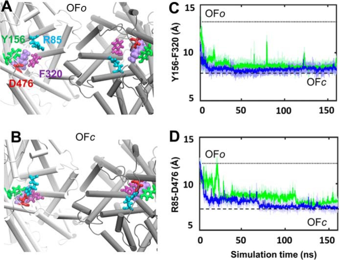
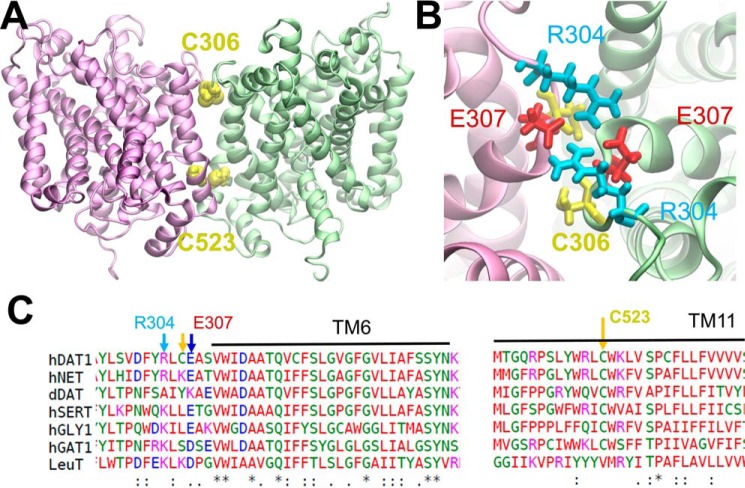
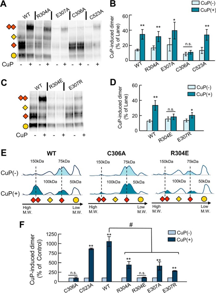
The human dopamine (DA) transporter (hDAT) is a key regulator of neurotransmission and a target for antidepressants and addictive drugs. Despite the recent resolution of dDAT structures from Drosophila melanogaster, complete understanding of its mechanism of function and even information on its biological assembly is lacking. The resolved dDAT structures are monomeric, but growing evidence suggests that hDAT might function as a multimer, and its oligomerization may be relevant to addictive drug effects. Here, using structure-based computations, we examined the possible mechanisms of hDAT dimerization and its dynamics in a lipid bilayer. Using a combination of site-directed mutagenesis, DA-uptake, and cross-linking experiments that exploited the capacity of Cys-306 to form intermonomeric disulfide bridges in the presence of an oxidizing agent, we tested the effects of mutations at transmembrane segment (TM) 6 and 12 helices in HEK293 cells. The most probable structural model for hDAT dimer suggested by computations and experiments differed from the dimeric structure resolved for the bacterial homolog, LeuT, presumably because of a kink at TM12 preventing favorable monomer packing. Instead, TM2, TM6, and TM11 line the dimer interface. Molecular dynamics simulations of the dimeric hDAT indicated that the two subunits tend to undergo cooperative structural changes, both on local (extracellular gate opening/closure) and global (transition between outward-facing and inward-facing states) scales. These observations suggest that hDAT transport properties may be allosterically modulated under conditions promoting dimerization. Our study provides critical insights into approaches for examining the oligomerization of neurotransmitter transporters and sheds light on their drug modulation.

Keywords: docking; dopamine transporter; molecular dynamics; mutagenesis; protein assembly.


Conflict of interest statement

The authors declare that they have no conflicts of interest with the contents of this article

Figures

Figure 1.
Figure 1.
The kink in the TM12 segment of DAT and its functional significance. Structural and sequence differences between LeuT from Aquifex aeolicus (A), dDAT from D. melanogaster (B), and hDAT (human) (C). There is a kink (dashed circle in B) in the TM12 segment of eukaryotic DATs near two serines conserved among the eukaryotic transporters. DAT has an additional C-terminal helix, with a glycine (G585 in hDAT) conserved among human paralogs. Notably, Ser-568 forms hydrogen bonding with Ser-483 in the TM10 segment. D, sequence alignment of TM12 in different members of transporters that share the LeuT fold. The sequences belong to human paralogs hDAT, hNET, hSERT, hGLY1, and hGAT1 and the fruit fly DAT (dDAT) shown in B. The last sequence is that of bacterial LeuT (shown in A). Selected residues conserved among all human paralogs are highlighted in yellow (also labeled in panel C). The kink-forming serines on TM12 are highlighted and enclosed in a dashed box. E and F, effect of mutations at TM12 kink-forming residues on DAT expression and DA uptake. E, surface expression of WT, G561A, S567F, S568L, and P573A hDAT. Transfected HEK293 cells were incubated with sulfo-NHS-SS-biotin and isolated with Neutravidin-agarose beads after cell lysis. For detection of DAT protein an anti-DAT (rat, Millipore MAB369) antibody was used. Densitometry analysis was performed with Image Lab (Bio-Rad) and SigmaPlot 12.5 (Systat Software). Bars represent the percentage of the mean value of the WT group (mean ± S.E.). F, transport activity. Uptake of [3H]DA was performed in HEK293 cells. DA accumulation was normalized with respect to the percentage of cell-surface expression (Surf. Exp.) for each mutant and expressed as the percentage of WT. The statistical analysis was performed with a two-tailed Student's t test versus WT group with an accepted significance level of p < 0.05 (**).
Figure 2.
Figure 2.
Difference in dimerization architecture between the known LeuT dimer and the computationally predicted hDAT dimer in the outward-facing conformation. A, LeuT dimer has interfacial TM segments TM9 (purple) and TM12 (lime). B, computationally predicted hDAT dimer makes interfacial contacts between TM6 (yellow), TM11 (orange), and TM2 (cyan) helices.
Figure 3.
Figure 3.
MD simulations of hDAT dimer dynamics, embedded into membrane lipids. A, typical MD set-up for hDAT dimer (colored in pink and green) in a lipid bilayer (yellow licorice format (55)) and solvated by 0.15 m NaCl (light blue). The bound substrate DA and the co-transported Na+ and Cl are represented by purple, yellow, and cyan spheres. B, alignment of 100-ns MD equilibrated hDAT dimer in the outward-facing (OF; orange ribbon diagram) and inward-facing (IF; cyan) state. The r.m.s.d. between the OF and IF dimers levels off at 3.4 ± 0.3 Å. C, comparison of helical orientations in the OF and IF states. For clarity, one monomer (in schematic format) taken from the dimer is shown for each state.
Figure 4.
Figure 4.
Time evolution of the interhelical distances. The opening/closure of the EC and IC vestibules is characterized by the respective interhelical distances of the EC-exposed TM1b-TM10 and the IC-exposed TM1a-TM6b, shown for both subunits, as labeled (see Refs. and 27). Results from four runs are presented: A, OF-1, in which the closure of the EC vestibule led to the OFc state in both subunits. B, OF-2, in which both subunits remained in the OFo state, as indicated by the separation between the EC-exposed TM1b and TM10 segments (light and dark blue) and the close proximity of the IC-exposed helical segments TM1a-TM6b (light and dark red). C, IF-1, in which the IC vestibule remained open in both subunits (see the red curves). D, IF-2, in which the initiation of a transition to the OF conformation could be observed, but the IC vestibule remained open in both subunits. The characteristic distances for TM1b-TM10 obtained in the hDAT dimer simulations are 20.0 ± 2.0 Å and 16.5 ± 0.5 Å in the OFo and OFc states, respectively. These values closely approximate those reported for hDAT monomer simulations (27). Distances are indicated by dotted and dashed lines in panels A and B. In the OF state the distance between the IC-exposed segments TM1a and TM6b remained 11.5 ± 1.0 Å, the same as that reported in the monomer simulations (27). In the IFo state, the IC vestibule is open for DA release when the distance between TM1a and TM6b is >15 Å in hDAT monomer (see Cheng and Bahar (27)).
Figure 5.
Figure 5.
Structural transition observed from the OFo to OFc in the hDAT dimer. Top view of A, the initial OFo state with the opening of two EC gates: Arg-85–Asp-476 and Tyr-156–Phe-320. B, closure of the EC gates in the OFc state; a snapshot around 160 ns was used. Shown is time evolution of the center of the mass distances between the two EC gating residue pairs (C) aromatic residues Tyr-156 and Phe-320 and (D) salt-bridge-forming residue pairs Arg-85 and Asp-476. Note that the closure of the gates occurred almost simultaneously in the two subunits (colored in green and blue).
Figure 6.
Figure 6.
Interfacial interactions. A, two interfacial cysteines between subunits I and II (colored pink and green) are likely to form cross-links between the dimers: Cys-306(I)- Cys-306(II) (superposed, not seen separately in the panel) and Cys-523(I)–Cys-523(II). They are within close distances (<11 Å) in both the OF and IF conformers. B, an interfacial salt bridge is likely to form between Glu-307 and Arg-304 in the IF conformation of the dimer. C, sequence alignment of the putative interfacial regions.
Figure 7.
Figure 7.
Oxidative cross-linking of DAT Cys-306 in TM6 hDAT mutants. A, representative blot of CuP cross-linking in intact cells WT and DAT mutants R304A, E307A, C306A, and C523A. Cross-linking reaction with 0.1 μm CuSO4 and 0.4 μm phenanthroline was performed for 5 min at room temperature. Detection of DAT protein was detected by Western blot using an anti-DAT (rat, Millipore MAB369) antibody. Different immunodetected DAT bands were detected in CuP non-treated (−) and treated lanes (+). Different transporter forms are indicated as follows: immature monomer (yellow circle), mature monomer (red rhombus), immature dimer (yellow rhombus), mature dimer (double red rhombus). B, quantification of the CuP-induced 150-kDa DAT abduct. Bars represent the intensity of the band as % of the total bands per lane. Densitometry analysis was performed with ImageLab (Bio-Rad) and SigmaPlot 12.5 (Systat Software). C and D, CuP cross-linking of WT, R304E, and E307R. E, the intensity lane profile (obtained with Image Lab Software) shows the relative position of the different DAT-forms for WT, C306A, and R304E. The shadowed peaks represent the mature forms (monomeric 75 kDa) and CuP-induced dimer (150 kDa). F, quantification of the CuP-induced dimers relative to the 150-kDa DAT band in the control group (CuP(−)) for each DAT construct. Bars represent the mean values (mean ± S.E.). *, p < 0.05; **, p < 0.01 for Student's t test; p value versus control group. # represents p < 0.05 analysis of variance one way; n.s., not significant.

References

    1. Amara S. G., and Sonders M. S. (1998) Neurotransmitter transporters as molecular targets for addictive drugs. Drug Alcohol Depend. 51, 87–96 - PubMed
    1. Reith M. (2002) Neurotransmitter transporters: Structure, Function, and Regulations. Humana, Totowa, NJ
    1. Vaughan R. A., and Foster J. D. (2013) Mechanisms of dopamine transporter regulation in normal and disease states. Trends. Pharmacol. Sci. 34, 489–496 - PMC - PubMed
    1. Jardetzky O. (1966) Simple allosteric model for membrane pumps. Nature 211, 969–970 - PubMed
    1. Sitte H. H., and Freissmuth M. (2015) Amphetamines, new psychoactive drugs, and the monoamine transporter cycle. Trends Pharmacol. Sci. 36, 41–50 - PMC - PubMed

Publication types

MeSH terms

Substances

LinkOut - more resources