. 2017 Nov;102(5):1159-1171.
doi: 10.1189/jlb.3HI1216-511RR. Epub 2017 Jun 5.

Frontline Science: Aspirin-triggered resolvin D1 controls herpes simplex virus-induced corneal immunopathology

Affiliations

Frontline Science: Aspirin-triggered resolvin D1 controls herpes simplex virus-induced corneal immunopathology

Naveen K Rajasagi et al. J Leukoc Biol. 2017 Nov.

Abstract

Stromal keratitis (SK) is a chronic immunopathological lesion of the eye, caused by HSV-1 infection, and a common cause of vision impairment in humans. The inflammatory lesions in the cornea are primarily caused by neutrophils with the active participation of CD4+ T cells. Therefore, the targeting of these immune cell types and their products represents a potentially valuable form of therapy to reduce the severity of disease. Resolvin D1 (RvD1) and its epimer aspirin-triggered RvD1 (AT-RvD1) are lipid mediators derived from docosahexaenoic acid (DHA) and were shown to promote resolution in several inflammatory disease models. In this report, we examined whether AT-RvD1 administration, begun before infection or at a later stage after ocular infection of mice with HSV-1, could control the severity of SK lesions. Treatment with AT-RvD1 significantly diminished the extent of corneal neovascularization and the severity of SK lesions. AT-RvD1-treated mice had fewer numbers of inflammatory cells that included neutrophils as well as Th1 and Th17 cells in the infected cornea. The mechanisms by which AT-RvD1 acts appear to be multiple. These include inhibitory effects on proinflammatory mediators, such as IL-1β, IL-6, IL-12, CXCL1, MCP-1, MIP-2, vascular endothelial growth factor (VEGF)-A, matrix metalloproteinase 9 (MMP-9), and proinflammatory miRNA, such as miR-155, miR-132, and miR-223, which are involved in SK pathogenesis and corneal neovascularization. In addition, AT-RvD1 attenuated STAT1, which plays an important role in Th1 cell differentiation and IFN-γ expression. These findings demonstrate that AT-RvD1 treatment could represent a useful strategy for the management of virus-induced immunopathological lesions.

Keywords: inflammation; resolvins; stromal keratitis.


Figures

Figure 1.
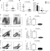
Figure 1.. Effect of prophylactic treatment with AT-RvD1 on HSV-induced ocular immunopathology.
Balb/c mice infected with 1 × 10⁵ PFU of HSV-1 RE were given AT-RvD1 topically, twice daily, starting from 1 d before infection until day 10 pi. (A and B) SK lesion severity and angiogenesis scores at day 15 pi are shown. Statistical significance was determined by Mann-Whitney test; n = 14 mice/group, as indicated in the scatter plots. (C) At the indicated time points, eyes of HSV-infected mice were swabbed with a sterile swab and assayed for infectious virus by standard plaque assay. The level of significance was determined by Student’s t test (unpaired). Error bars represent means ± sem (n = 8 eye swabs/group). (D–I) The immune parameters were analyzed at the termination of the experiment (day 15 pi). Representative dot plots show percentage of (D) leukocytes (CD45+), (F) neutrophils (CD11B+Ly6G+), and (H) CD4+ T cells in the inflamed cornea of control and AT-RvD1-treated animals at day 15 pi. The average number of (E) CD45+ cells, (G) CD11B+Ly6G+, and (I) CD4+ T cells per cornea at indicated time points is shown. The level of significance was determined by Mann-Whitney test. Error bars represent means ± sem; n = 7 corneas per group. Experiments were repeated at least 2 times. P < 0.05 was regarded as a significant difference between groups. SSC, Side-scatter.
Figure 2.
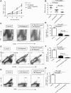
Figure 2.. Topical administration of AT-RvD1, started at day 6 pi, decreases SK severity in HSV-1-infected mice.
Balb/c mice infected with 1 × 10⁵ PFU of HSV-1 RE were given AT-RvD1 topically, twice daily, starting from day 6 until day 13 pi. The disease severity was examined on different days pi. (A–C) Disease progression (until day 15 pi), SK lesion severity, and angiogenesis at day 15 pi are shown. The level of significance was determined by Mann-Whitney test. Error bars represent means ± sem; n = 16 mice/group, as indicated in the scatter plots. (D–K) The immune parameters were analyzed at the termination of the experiment (day 15 pi). Representative dot plots show percentage of (D) leukocytes (CD45+), (F) neutrophils (CD11B+Ly6G+), (H) CD4+ T cells, and (J) CD31+ cells in the inflamed cornea of control and AT-RvD1-treated animals at day 15 pi. The average number of (E) CD45+ cells, (G) CD11B+Ly6G+, (I) CD4+ T cells, and (K) CD31+ cells per cornea at indicated time points is shown. The level of significance was determined by Mann-Whitney test. Error bars represent means ± sem; n = 5 corneas per group. Experiments were repeated at least 3 times. P < 0.05 was regarded as a significant difference between groups.
Figure 3.
Figure 3.. Topical administration of AT-RvD1, started at day 6 pi, reduced Th1 and Th17 cell responses in the corneas of HSV-1-infected mice.
Balb/c mice infected with 1 × 10⁵ PFU of HSV-1 RE were given AT-RvD1 topically, twice daily, starting from day 6 until day 13 pi. Representative plots show percentage of CD4+ T cells producing (A) IFN-γ, (C) IL-2, or (E) IL-17 in the corneas of control and AT-RvD1-treated animals following stimulation with CD3/CD28. Plots shown were gated on CD4+ T cells. The average number of CD4 cells producing (B) IFN-γ, (D) IL-2, and (F) IL-17 in the cornea at day 15 pi is shown. The level of significance was determined by Mann-Whitney test. Error bars represent means ± sem; n = 5 corneas per group. Experiments were repeated at least 3 times. P < 0.05 was regarded as a significant difference between groups.
Figure 4.
Figure 4.. Effect of prophylactic treatment with AT-RvD1 during acute phase of HSV-1 corneal Infection.
Balb/c mice infected with 1 × 10⁵ PFU of HSV-1 RE were given AT-RvD1 topically, twice daily, starting either from 1 d before infection until day 6 pi (acute phase) or 1 d before infection until day 14 pi. (A and B) HSK disease progression and SK lesion severity at day 15 pi are shown. Statistical significance was determined by Mann-Whitney test; n = 15 mice/group, as indicated in the scatter plots. (C–H) The immune parameters were analyzed at the termination of the experiment (day 15 pi). Representative dot plots show percentage of (C) leukocytes (CD45+), (D) neutrophils (CD11B+Ly6G+), and (E) CD4+ T cells in the inflamed cornea of control and AT-RvD1-treated animals at day 15 pi. The average number of (F) CD45+ cells, (G) CD11B+Ly6G+, and (H) CD4+ T cells per cornea at indicated time points is shown. The level of significance was determined by Mann-Whitney test. Error bars represent means ± sem; n = 5 corneas per group. Data shown are from 2 independent experiments. P < 0.05 was regarded as a significant difference between groups.
Figure 5.
Figure 5.. Topical administration of AT-RvD1, started at day 6 pi, dampened the expression of proinflammatory mediators in the corneas of HSV-1-infected mice.
Balb/c mice infected with 1 × 10⁵ PFU of HSV-1 RE were given AT-RvD1 topically, twice daily, starting from day 6 until day 13 pi. Mice were euthanized at day 15 pi, and the expression levels of cytokines, chemokines, and angiogenic factors were measured in 6 different pooled corneal samples, each consisting of 4 corneas/group in control and AT-RvD1-treated animals using qRT-PCR. Expression levels were the following: (A) IL-1β, (B) IL-6, (C) IL-12, (D) CXCL1, (E) MCP-1, (F) MIP-2, (G) VEGF-A, and (H) MMP-9 in corneal samples obtained from control and AT-RvD1-treated animals. The level of significance was determined by Mann-Whitney test. Error bars represent means ± sem; n = 6 samples per group. P < 0.05 was regarded as a significant difference between groups.
Figure 6.
Figure 6.. Topical AT-RvD1 administration reduced the expression of proinflammatory miRNA in the corneas of HSV-1-infected mice.
Balb/c mice infected with 1 × 10⁵ PFU of HSV-1 RE were given AT-RvD1 topically, twice daily, starting from day 6 until day 13. Mice were euthanized at day 15 pi, and expression levels of miRNA were determined in 4 different pooled corneal samples, each consisting of 4 corneas/group in control and AT-RvD1-treated animals using qRT-PCR. Expression levels were the following: (A) miR-155, (B) miR-132, and (C) miR-223 in corneal samples obtained from control and AT-RvD1-treated animals. The level of significance was determined using Mann-Whitney test. Error bars represent means ± sem; n = 4 samples per group. P < 0.05 was regarded as a significant difference between groups.
Figure 7.
Figure 7.. AT-RvD1 can diminish IFN-γ production in CD4+ T cells activated in vitro and reduced pSTAT1.
(A and B) Balb/c mice infected with 1 × 10⁵ PFU of HSV-1 RE were euthanized on day 15 pi, spleen cells were isolated, and in vitro experiments were performed, as mentioned in Materials and Methods. (A) Representative dot plots show percentage of CD4+ T cells producing IFN-γ in the untreated control and AT-RvD1-treated samples following stimulation with CD3/CD28. Plots shown were gated on CD4+ T cells. (B) Proportion of CD4+ cells producing IFN-γ in the control and samples treated with different concentrations of AT-RvD1 (100, 500, and 1000 nM). The level of significance was determined by Mann-Whitney test comparing control with different concentration of AT-RvD1. Error bars represent means ± sem; n = 4 per group. Experiments were repeated at least 3 times. P < 0.05 was regarded as a significant difference between groups. (C and D) Spleen cells were obtained from naive Balb/c mice, and in vitro experiments were performed to measure pSTAT1 in CD4+ T cells using flow cytometry, as mentioned in Materials and Methods. (C) Representative FACS plots show percentage of CD4+ pSTAT1+ T cells in unstimulated and control and AT-RvD1-treated samples following IFN-γ stimulation. Plots shown were gated on CD4+ T cells. (D) Proportion of CD4+ pSTAT1+ T cells in the control and samples treated with different concentrations of AT-RvD1 (100, 500, and 1000 nM). The level of significance was determined by Mann-Whitney test comparing control with different concentration of AT-RvD1. Error bars represent means ± sem; n = 4 per group. Experiments were repeated at least 2 times. P < 0.05 was regarded as a significant difference between groups.
Figure 8.
Figure 8.. Depletion of CD4 T cells diminished the development of SK lesions.
Control and CD4-depleted Balb/c mice were infected with 1 × 10⁵ PFU of HSV-1 RE. CD4-depleted mice were left untreated or treated with AT-RvD1 topically, twice daily, starting from day 6 until day 13 pi. (A) The disease progression was examined on different days pi. (B) SK lesion severity at day 15 pi. The level of significance was determined by Mann-Whitney test. Error bars represent means ± sem; n = 15 mice/group, as indicated in the scatter plots. (C–H) Corneal infiltration was examined at the termination of the experiment (day 15 pi). Representative dot plots show percentage of (C) leukocytes (CD45+), (E) neutrophils (CD11B+Ly6G+), and (G) CD4+ T cells in the cornea of control, CD-depleted, and CD4 depleted/AT-RvD1-treated animals at day 15 pi. The average number of (D) CD45+ cells, (F) CD11B+Ly6G+, and (H) CD4+ T cells per cornea at day 15 pi is shown. The data are pooled from 2 experiments, with n = 5 corneas per group. The level of significance was determined by Mann-Whitney test. Error bars represent means ± sem. P < 0.05 was regarded as a significant difference between groups.

Comment in

Similar articles

Cited by

References

    1. Liesegang T. J. (2001) Herpes simplex virus epidemiology and ocular importance. Cornea 20, 1–13. - PubMed
    1. Rowe A. M., St Leger A. J., Jeon S., Dhaliwal D. K., Knickelbein J. E., Hendricks R. L. (2013) Herpes keratitis. Prog. Retin. Eye Res. 32, 88–101. - PMC - PubMed
    1. Deshpande S., Banerjee K., Biswas P. S., Rouse B. T. (2004) Herpetic eye disease: immunopathogenesis and therapeutic measures. Expert Rev. Mol. Med. 6, 1–14. - PubMed
    1. McGhee C. N., Dean S., Danesh-Meyer H. (2002) Locally administered ocular corticosteroids: benefits and risks. Drug Saf. 25, 33–55. - PubMed
    1. Knickelbein J. E., Hendricks R. L., Charukamnoetkanok P. (2009) Management of herpes simplex virus stromal keratitis: an evidence-based review. Surv. Ophthalmol. 54, 226–234. - PubMed

Publication types

MeSH terms