. 2017 Jun 27;114(26):E5158-E5166.
doi: 10.1073/pnas.1621076114. Epub 2017 Jun 5.

Deubiquitinating enzyme VCIP135 dictates the duration of botulinum neurotoxin type A intoxication

Affiliations

Deubiquitinating enzyme VCIP135 dictates the duration of botulinum neurotoxin type A intoxication

Yien Che Tsai et al. Proc Natl Acad Sci U S A. .

Abstract

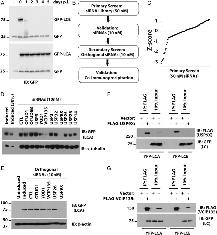
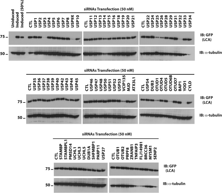
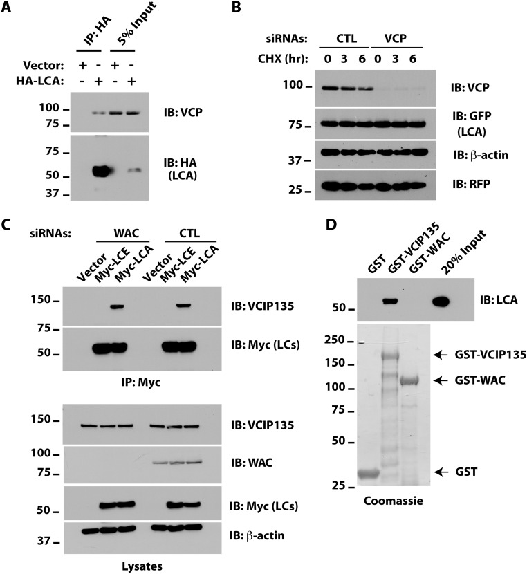
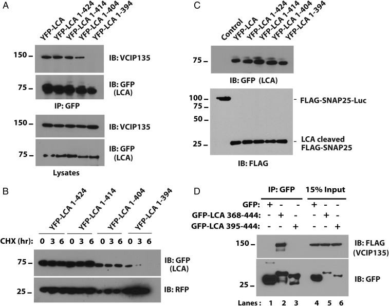
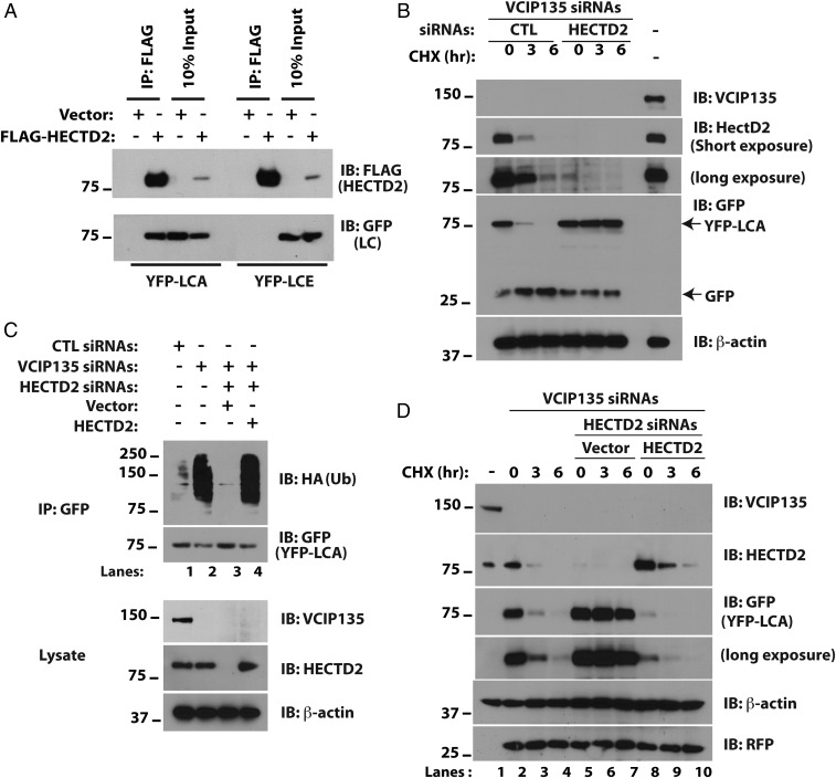
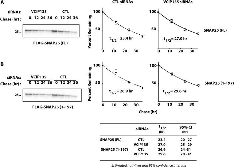
Botulism is characterized by flaccid paralysis, which can be caused by intoxication with any of the seven known serotypes of botulinum neurotoxin (BoNT), all of which disrupt synaptic transmission by endoproteolytic cleavage of SNARE proteins. BoNT serotype A (BoNT/A) has the most prolonged or persistent effects, which can last several months, and exerts its effects by specifically cleaving and inactivating SNAP25. A major factor contributing to the persistence of intoxication is the long half-life of the catalytic light chain, which remains enzymatically active months after entry into cells. Here we report that BoNT/A catalytic light chain binds to, and is a substrate for, the ubiquitin ligase HECTD2. However, the light chain evades proteasomal degradation by the dominant effect of a deubiquitinating enzyme, VCIP135/VCPIP1. This deubiquitinating enzyme binds BoNT/A light chain directly, with the two associating in cells through the C-terminal 77 amino acids of the light chain protease. The development of specific DUB inhibitors, together with inhibitors of BoNT/A proteolytic activity, may be useful for reducing the morbidity and public health costs associated with BoNT/A intoxication and could have potential biodefense implications.

Keywords: USP9X; motoneuron; synaptic transmission; synaptosomal-associated protein 25; toxin persistence.


Conflict of interest statement

Conflict of interest statement: G.A.O. has a financial interest in Synaptic Research LLC, which is developing therapeutics for BoNT intoxication, including the delivery of designer ubiquitin ligases.

Figures

Fig. 1.
Fig. 1.
Identification of DUBs that stabilize BoNT/A catalytic LC. (A) Cell culture model for persistence of LCA. Flp-in/T-REX/ 293 cells were induced with doxycycline for 16 h to express GFP-LCE and GFP-LCA. The persistence of GFP-LCs was monitored following doxycycline withdrawal (postinduction, p.i.). GFP served as the loading and transfection control. (B) Algorithm for siRNA library screening. (C) The z score for primary screen was computed based on median and SD of the sample (Materials and Methods). (D) Validation of potential hits from primary screen with low concentrations (10 nM) of the same siRNAs compared with control (CTL) siRNAs. The lane labeled “Induced (30%)” contains 30% of the cell lysate compared with other lanes. (E) Secondary screen with low concentrations (10 nM) of orthogonal siRNAs from a different vendor. α-tubulin and β-actin served as loading controls in D and E, respectively. (F and G) Coimmunoprecipitation assays to evaluate interactions of LCA with USP9X (F) or VCIP135 (G).
Fig. S1.
Fig. S1.
Identification of DUBs that stabilize BoNT/A catalytic LC. Flp-in/T-REX/293 cells were induced with doxycycline for 16 h to express GFP-LCA. On the following day, cells were washed, trypsinized, replated, and allowed to recover overnight. The primary screen was carried out with 50 nM siRNAs and cells harvested 3 d posttransfection. Levels of LCA were monitored by Western blotting.
Fig. S2.
Fig. S2.
Different pathways for LCA degradation in USP9X- and VCIP135-depleted cells. (A) HEK293 cells were transfected with YFP-LCA, and the indicated siRNAs and degradation of YFP-LCA (recognized by GFP antibody) were assessed by CHX chase and Western blotting. (B) M17 cells transfected with VCIP135 siRNAs and YFP-LCA were treated with the indicated inhibitors with or without CHX for 6 h and YFP-LCA monitored. (C) M17 cells were transfected with YFP-LCA and either VCIP135 or USP9X siRNAs with or without ATG siRNAs and degradation of YFP-LCA assessed. RFP was included as a transfection efficiency control and as a loading control along with β-actin.
Fig. 2.
Fig. 2.
The catalytic activity of VCIP135 is necessary for stabilizing LCA. (A) M17 cells were transfected with YFP-LCA and the indicated siRNAs and degradation of YFP-LCA (recognized by GFP antibody) assessed by CHX chase and Western blotting. (B) Cells transfected with VCIP135 siRNAs and YFP-LCA were treated with CHX and the indicated inhibitors or vehicle control (DMSO) for 6 h. RFP served as a transfection and loading control. (C) Cells were transfected with VCIP135 siRNAs and subsequently transfected with YFP-LCA and the indicated VCIP135 constructs. (D) Cells were transfected with YFP-LCA and the indicated siRNAs.
Fig. S3.
Fig. S3.
Different pathways for LCA degradation in USP9X- and VCIP135-depleted cells. (A) M17 cells were transfected with HA-tagged LCA. After 48 h, cells were harvested and HA-LCA immunoprecipitates were immunoblotted as indicated. (B) Cells were transfected with YFP-LCA and either CTL or VCP siRNAs. Degradation of YFP-LCA was assessed by CHX chase. (C) Cells were transfected with CTL or WAC siRNAs. After 18 h, cells were transfected with plasmids encoding either Myc-LCA or Myc-LCE. Myc-LC proteins were immunoprecipitated with Myc agarose and samples immunoblotted as indicated. (D) E. coli lysates containing ∼0.2 μg 6×His-LCA and 2 μg of GST fusion proteins were incubated with Glutathione Sepharose 4B for 2 h at 4 °C and LCA binding assessed by immunoblotting. The relative amounts of GST fusion proteins used in the binding assay were assessed by enrichment using Glutathione Sepharose 4B and Coomassie staining and are shown below. Bands beneath the full-length proteins in the Coomassie gel represent degradation products and/or products of incomplete translation.
Fig. 3.
Fig. 3.
The C terminus of LCA interacts with VCIP135. (A) HEK293 cells were transfected with the indicated C-terminal deletion mutants of YFP-LCA. YFP-LCA mutants were immunoprecipitated with GFP antibody and immunoblotted for VCIP135. (B) HEK293 cells were transfected with deletion mutants as in A, and degradation of YFP-LCAs was assessed. (C) HEK293 cells were transfected with FLAG-SNAP25-luciferase and the indicated YFP-LCAs. After 36 h, the proteolytic activity of the truncation mutants was assessed by evaluating cleavage of FLAG-SNAP25-luciferase using FLAG antibody. (D) HEK293 cells were transfected with the C-terminal 77 or 50 residues of LCA fused to GFP. After 30 h, GFP-LCA fusions were immunoprecipitated with GFP antibody and coimmunoprecipitated VCIP135 assessed.
Fig. 4.
Fig. 4.
HECTD2 promotes the degradation of LCA. (A) HEK293 cells were transfected with plasmids encoding FLAG-HECTD2 and YFP-LCA as indicated. After 36 h, cells were treated with MG132 for 8 h to inhibit the proteasome and lysed in Triton X-100 buffer. (B) M17 cells depleted of VCIP135 were transfected with YFP-LCA and either CTL or HECTD2 siRNAs. After 48 h, cells were treated with CHX and loss of YFP-LCA assessed. Cotransfected GFP was used to monitor transfection efficiency. (C) M17 cells were transfected with YFP-LCA, HA-ubiquitin, and the indicated siRNAs and plasmids. After 72 h, cells were treated with MG132 for 8 h followed by cell lysis, immunoprecipitation of YFP-LCA, and immunoblotting for HA-ubiquitin. (D) Cells were transfected with the indicated siRNAs and replated for overnight culture. After 16 h, cells were transfected with either vector or plasmid expressing HECTD2 or were not subject to further transfection (lanes 1–4).
Fig. S4.
Fig. S4.
Identification of a ubiquitin ligase for BoNT/A catalytic LC. (A) Schematic of the screen. (B) Western blots of siRNA screen of HECT domain ubiquitin ligases identifying HECTD2 as a putative ubiquitin ligase for LCA. The lane labeled “CTL (50%)” contains 50% of the cell lysate compared with other lanes. (C) E. coli lysates containing ∼0.2 μg 6×His-LCA and 2 μg of GST fusion proteins were assessed as in S3D. (D) Cells were transfected with FLAG-HECTD2 and full-length YFP-LCA or the indicated deletion mutants. FLAG-HECTD2 was immunoprecipitated with M2 agarose and coimmunoprecipitated YFP proteins assessed. (E) Cells were transfected with the indicated plasmids. Immunoprecipitation for EE-tagged HECTD2 was followed by immunoblotting as indicated.
Fig. S5.
Fig. S5.
Effect of VCIP135 depletion on turnover of SNAP25. M17 cells were transfected with CTL or VCIP135 siRNAs 18 h before transfection with (A) FLAG-SNAP25 or (B) FLAG-SNAP251–197. Turnover of SNAP25 proteins was assessed by pulse-chase metabolic labeling. Representative data shown on the Left and summarized on the Right (mean ± SD; n = 3). The half-lives of full-length SNAP25 and SNAP251–197 were ∼23 and 27 h, respectively, comparable to their reported half-lives of 24 h in cultured neurons (36). Importantly, depletion of VCIP135 had little effect on the measured half-lives for SNAP25 (t1/2 = 27 h) or SNAP251–197 (t1/2 = 30 h); P > 0.99 by Bonferroni’s multiple comparison tests.
Fig. 5.
Fig. 5.
Depletion of VCIP135 accelerates recovery of full-length SNAP25 from BoNT. (A) Motoneurons derived from mouse embryonic stem cells were treated with BoNT/A holotoxin. After 20 h, cells were washed with media to remove residual toxin and transfected with either CTL or VCIP135 siRNAs. Cell lysates were collected at the indicated days postintoxication and levels of full-length and cleaved SNAP25 monitored by Western blotting, which migrated as expected at 25 and 24 kDa, respectively. (B) Semiquantitative analysis of the recovery of full-length SNAP25 as a percentage of total SNAP25. Data were shown as mean ± SEM (n = 3). **P < 0.01 based on Bonferroni’s multiple comparison tests (Table 1). Note that the initial knockdown of VCIP135 in experiment 2 was ineffective; regardless, the corresponding VCIP135 data were included in this analysis.

Similar articles

Cited by

References

    1. Simpson LL. Identification of the characteristics that underlie botulinum toxin potency: Implications for designing novel drugs. Biochimie. 2000;82:943–953. - PubMed
    1. Habermann E, Dreyer F. Clostridial neurotoxins: Handling and action at the cellular and molecular level. Curr Top Microbiol Immunol. 1986;129:93–179. - PubMed
    1. Johnson EA, Montecucco C. Botulism. Handb Clin Neurol. 2008;91:333–368. - PubMed
    1. Schiavo G, Matteoli M, Montecucco C. Neurotoxins affecting neuroexocytosis. Physiol Rev. 2000;80:717–766. - PubMed
    1. Montecucco C, Schiavo G. Structure and function of tetanus and botulinum neurotoxins. Q Rev Biophys. 1995;28:423–472. - PubMed

Publication types

MeSH terms

LinkOut - more resources