. 2017 Jun 20;114(25):6557-6562.
doi: 10.1073/pnas.1619660114. Epub 2017 Jun 5.

Crystal structures of the Burkholderia multivorans hopanoid transporter HpnN

Affiliations

Crystal structures of the Burkholderia multivorans hopanoid transporter HpnN

Nitin Kumar et al. Proc Natl Acad Sci U S A. .

Abstract

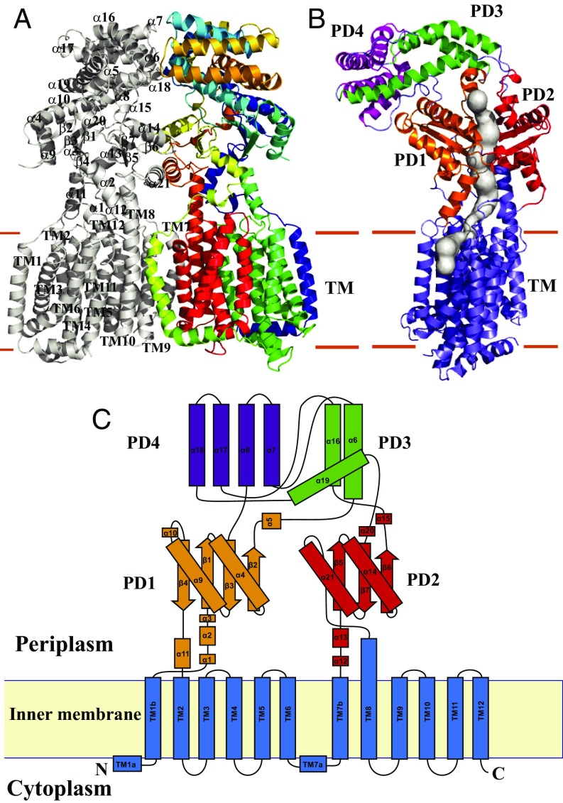
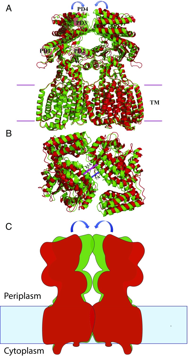
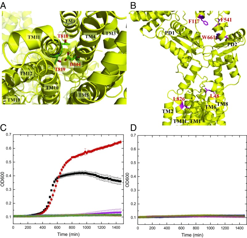
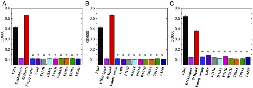
Strains of the Burkholderia cepacia complex (Bcc) are Gram-negative opportunisitic bacteria that are capable of causing serious diseases, mainly in immunocompromised individuals. Bcc pathogens are intrinsically resistant to multiple antibiotics, including β-lactams, aminoglycosides, fluoroquinolones, and polymyxins. They are major pathogens in patients with cystic fibrosis (CF) and can cause severe necrotizing pneumonia, which is often fatal. Hopanoid biosynthesis is one of the major mechanisms involved in multiple antimicrobial resistance of Bcc pathogens. The hpnN gene of B. multivorans encodes an integral membrane protein of the HpnN family of transporters, which is responsible for shuttling hopanoids to the outer membrane. Here, we report crystal structures of B. multivorans HpnN, revealing a dimeric molecule with an overall butterfly shape. Each subunit of the transporter contains 12 transmembrane helices and two periplasmic loops that suggest a plausible pathway for substrate transport. Further analyses indicate that HpnN is capable of shuttling hopanoid virulence factors from the outer leaflet of the inner membrane to the periplasm. Taken together, our data suggest that the HpnN transporter is critical for multidrug resistance and cell wall remodeling in Burkholderia.

Keywords: Burkholderia multivorans; HpnN transporter; cell wall remodeling; hopanoid transport; multidrug resistance.


Conflict of interest statement

The authors declare no conflict of interest.

Figures

Fig. 1.
Fig. 1.
Structure of the B. mulitvorans HpnN transporter. (A) Ribbon diagram of a dimer of HpnN viewed in the membrane plane. The Right subunit of the dimer is colored using a rainbow gradient from the N terminus (blue) to the C terminus (red), whereas the Left subunit is colored gray. Overall, the HpnN dimer forms a butterfly-shaped structure. (B) Each subunit of the HpnN transport forms a channel (colored gray) spanning the outer leaflet of the inner membrane and up to the periplasmic domain. This figure depicts the Left subunit of the form I structure of the HpnN dimer. The orientation of this HpnN subunit has been rotated by 60° counterclockwise, based on the vertical C2 symmetry axis of the HpnN dimer, compared with the orientation of A. This channel was calculated using the program CAVER (loschmidt.chemi.muni.cz/caver). The transmembrane helices are colored slate. PD1–PD4 are colored orange, red, green, and magenta, respectively. (C) Secondary structural topology of the HpnN monomer. The topology was constructed based on the crystal structure of HpnN. The TM domain, PD1–PD4 are colored blue, orange, red, green, and purple, respectively.
Fig. 2.
Fig. 2.
Channel in the HpnN transporter. (A) Channel formed by each subunit of the form I structure of HpnN. Each channel is colored gray. The Left and Right subunits of HpnN are colored yellow and red, respectively. (B) Channel formed by each subunit of the form II structure of HpnN. Each channel is colored gray. The Left and Right subunits of HpnN are colored green and orange, respectively. The calculations were done using the program CAVER (loschmidt.chemi.muni.cz/caver). (C) Schematic representation illustrating the channel is open in each subunit of the form I structure of HpnN. The HpnN transporter may use this open channel to shuttle hopanoid molecules to the outer membrane. (D) Schematic representation illustrating the channel is closed in each subunit of the form II structure of HpnN. In this conformation, hopanoid molecules may not be able to pass through the channel.
Fig. 3.
Fig. 3.
Structural comparison of forms I and II of the HpnN transporter. (A) Superimposition of the dimeric structures of forms I and II (green, form I; red, form II). For clarity, only the Left subunit, PD1–PD4, is labeled. Each arrow indicates a rigid-body swinging motion of the periplasmic domain of each monomer, allowing it to come closer to the next subunit within the dimer. (B) Superimposition of the dimeric structures of forms I and II (green, form I; red, form II) viewed from the periplasmic side. This view depicts the two monomers of the form I structure as at least 6 Å closer to each other at the dimer interface compared with those of the form II structure. For clarity, the transmembrane domain of the HpnN transporter is not included in this figure. (C) Schematic representation illustrating the rigid-body swinging motion of the periplasmic domains of dimeric HpnN. Each arrow indicates a rigid body swinging motion of the periplasmic domain of each monomer. The swinging motion is capable of converting the conformations between form I (green) and form II (red).
Fig. 4.
Fig. 4.
Important conserved amino acids of HpnN. (A) Ion pairs in the transmembrane domain viewed from the cytoplasmic side. Residues D344 of TM4, and T818 and T819 of TM11 that form ion pairs, which may play an important role in proton translocation, are in green sticks. (B) Side view of a protomer of HpnN that forms a channel. Residues L48, F117, F541, W661, and L826, which line the wall of the channel are in magenta sticks. (C) Time course of the growth of B. thailandensis E264∆hpnN cells harboring mutants of the proton relay network. Cells expressing the mutant transporter D344Y, T818A, or T819A could not grow in liquid LB in the presence of 5 μg/mL chloramphenicol (black, E264 cells; red, E264∆hpnN/pHERD20Ωbt_hpnN cells expressing HpnN; magneta, E264∆hpnN cells; blue, E264∆hpnN/pHERD20 cells; cyan, cells expressing D344Y; orange, T818A; and green, T819A). Error bars represent SD (n = 3). (D) Time course of the growth of B. thailandensis E264∆hpnN cells harboring mutant transporters L48F, F117R, F541R, W661H, and L826F. Growth of cells expressing these mutant transporters was severely attenuated in liquid LB supplemented with 5 μg/mL chloramphenicol (deep purple, L48F; gray, F117R; hot pink, F541R; smudge, W661H; and dark blue, L826F). Growth of E264 cells, E264∆hpnN cells, E264∆hpnN/pHERD20Ωbt_hpnN cells expressing B. thailandensis HpnN, and E264∆hpnN/pHERD20 cells carrying the empty vector is shown in A. Error bars represent SD (n = 3).
Fig. 5.
Fig. 5.
Growth of cells in the presence of antibiotics. B. thailandensis E264∆hpnN cells expressing the mutant transporter L48F, F117R, D344Y, F541R, W661H, T818A, T819A, or L826F were retarded in growth in liquid LB supplemented with (A) 5 μg/mL chloramphenicol, (B) 1 μg/mL novobiocin, or (C) 1 mg/mL polymyxin B compared with cells expressing wild-type B. thailandensis HpnN. Error bars represent SD (n = 3). *Values of E264∆hpnN/pHERD20 and E264∆hpnN cells expressing the mutant transporters that are significantly lower than that of E264∆hpnN/pHERD20Ωbt_hpnN expressing wild-type HpnN (P < 6 × 10⁻⁶ for A, P < 2 × 10⁻⁶ for B, and P < 7 × 10⁻⁵ for C; Student’s t test).

References

    1. Mahenthiralingam E, Urban TA, Goldberg JB. The multifarious, multireplicon Burkholderia cepacia complex. Nat Rev Microbiol. 2005;3:144–156. - PubMed
    1. Sajjan U, et al. Immunolocalisation of Burkholderia cepacia in the lungs of cystic fibrosis patients. J Med Microbiol. 2001;50:535–546. - PubMed
    1. Ledson MJ, Gallagher MJ, Corkill JE, Hart CA, Walshaw MJ. Cross infection between cystic fibrosis patients colonised with Burkholderia cepacia. Thorax. 1998;53:432–436. - PMC - PubMed
    1. Zlosnik JE, et al. Burkholderia species infections in patients with cystic fibrosis in British Columbia, Canada. 30 years’ experience. Ann Am Thorac Soc. 2015;12:70–78. - PubMed
    1. Drevinek P, Mahenthiralingam E. Burkholderia cenocepacia in cystic fibrosis: Epidemiology and molecular mechanisms of virulence. Clin Microbiol Infect. 2010;16:821–830. - PubMed

Publication types

MeSH terms

Substances

LinkOut - more resources