

Published Erratum
doi: 10.1073/pnas.1708137114.
Epub 2017 Jun 5.
Correction for Ferrari et al., Hypoxia treatment reverses neurodegenerative disease in a mouse model of Leigh syndrome
- PMID: 28584118
- PMCID: PMC5474776
- DOI: 10.1073/pnas.1708137114
Item in Clipboard
Published Erratum
Correction for Ferrari et al., Hypoxia treatment reverses neurodegenerative disease in a mouse model of Leigh syndrome
Proc Natl Acad Sci U S A.
.
No abstract available
Figures
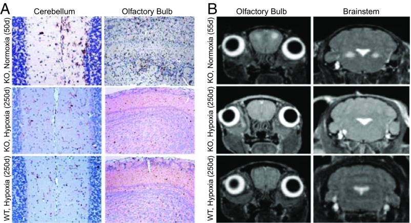
Absence of neurodegenerative pathology in 250-d-old hypoxia-treated Ndufs4 KO mice. (A) Representative images with staining for the microglial activation marker Iba-1 (n = 3). Normoxic KO mice at 50 d show a significant inflammatory response in the cerebellum and OB. Analogous images in 250-d-old hypoxic KO mice and WT mice do not show brain inflammation. (B) Axial head MRI images showing bilateral, symmetrical hyperintense lesions in the OB (Left) and brainstem (Right) of normoxic-breathing KO mice at 55 d. These lesions were not present in hypoxic KO mice at 250 d.
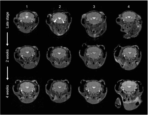
Breathing 11% O2 in late-stage neurological disease reverses the radiographic lesions of Ndufs4 mice seen on MRI. Four Ndufs4 mice were breathing 21% O2 until they developed late-stage neurological disease (55 d). They underwent MRI to document bilateral lesions in the vestibular nuclei (Upper, white arrow). Subsequently they commenced breathing 11% oxygen. The mice were scanned again at 2 wk and 4 wk of hypoxic breathing (Middle and Lower, respectively). Neurologic lesions disappeared by 4 wk of hypoxic breathing. The section of the fourth ventricle shown at the center of the brainstem appeared enlarged in the early scans, suggesting parenchymal atrophy. After 4 wk of treatment, this area appeared to be reduced in size, and morphological relationships were restored.
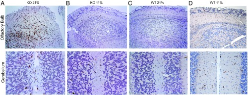
Breathing 11% O2 in late-stage neurological disease reverses pathological inflammation in the brains of Ndufs4 KO mice. Representative images with Iba-1 staining of the OB and cerebellum (CB) in KO mice and WT controls (n = 3 per group). Iba-1 is a marker of inflammation in the brain, indicative of microglial activation. Images demonstrate the reversibility of the neuropathological pattern by breathing 11% O2 at the late stage of disease (55 d). (A) KO mice breathing 21% O2. (B) KO mice breathing 21% O2 up to 55d and then breathing11% O2 (to 160d). MRI-demonstrated reversal of the lesions reported in Fig. 8 was observed in these same mice. (C) Normoxic WT controls. (D) Hypoxic WT controls.
Erratum for
-
Hypoxia treatment reverses neurodegenerative disease in a mouse model of Leigh syndrome.Proc Natl Acad Sci U S A. 2017 May 23;114(21):E4241-E4250. doi: 10.1073/pnas.1621511114. Epub 2017 May 8. Proc Natl Acad Sci U S A. 2017. PMID: 28483998 Free PMC article.
Publication types
LinkOut - more resources
Full Text Sources
Other Literature Sources
