. 2017 Jul 19;85(8):e00321-17.
doi: 10.1128/IAI.00321-17. Print 2017 Aug.

Chlamydia muridarum with Mutations in Chromosomal Genes tc0237 and/or tc0668 Is Deficient in Colonizing the Mouse Gastrointestinal Tract

Affiliations

Chlamydia muridarum with Mutations in Chromosomal Genes tc0237 and/or tc0668 Is Deficient in Colonizing the Mouse Gastrointestinal Tract

Lili Shao et al. Infect Immun. .

Abstract

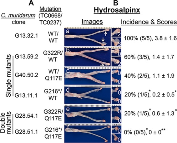
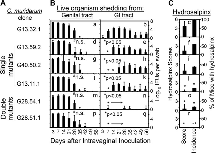
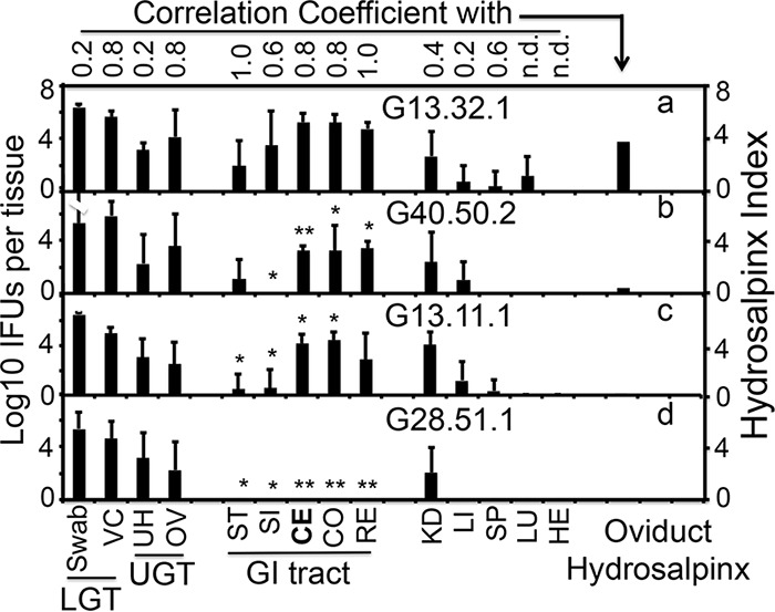
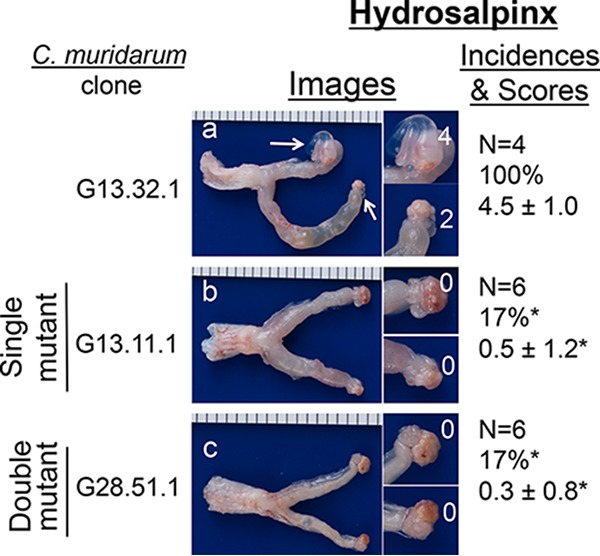
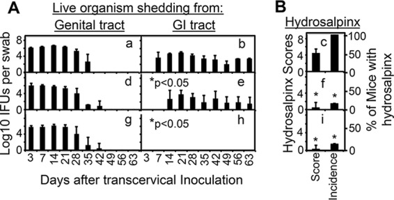
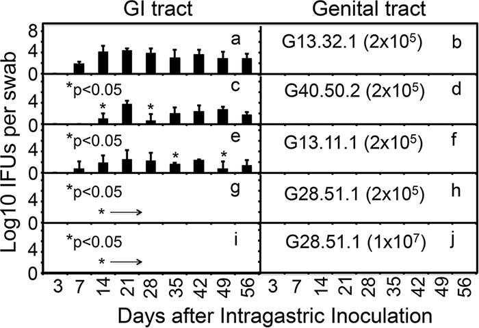
Chlamydiae colonize the gastrointestinal tracts of both animals and humans. However, their medical significance remains unknown. We have previously shown that wild-type Chlamydia muridarum spreads to and establishes stable colonization of the gastrointestinal tract following intravaginal inoculation. In the present study, we found that C. muridarum with mutations in chromosomal genes tc0237 and/or tc0668 was defective in spreading to the mouse gastrointestinal tract, which correlated with its attenuated pathogenicity in the upper genital tract. This correlation was more consistent than that of chlamydial pathogenicity with ascending infection in the genital tract, since attenuated C. muridarum spread significantly less to the gastrointestinal tract but maintained robust ascending infection of the upper genital tract. Transcervical inoculation further confirmed the correlation between C. muridarum spreading to the gastrointestinal tract and its pathogenicity in the upper genital tract. Finally, defective spreading of C. muridarum mutants was due to their inability to colonize the gastrointestinal tract since intragastric inoculation did not rescue the mutants' colonization. Thus, promoting C. muridarum colonization of the gastrointestinal tract may represent a primary function of the TC0237 and TC0668 proteins. Correlation of chlamydial colonization of the gastrointestinal tract with chlamydial pathogenicity in the upper genital tract suggests a potential role for gastrointestinal chlamydiae in genital tract pathogenicity.

Keywords: Chlamydia muridarum; TC0237; TC0668; attenuation; chlamydia; gut colonization; hydrosalpinx; mutants; mutations in tc0668 or tc0237; pathogenicity.


Figures

FIG 1
FIG 1
Ability of C. muridarum with or without mutations in tc0668 and/or tc0237 to induce hydrosalpinx in C3H/HeJ mice following intravaginal inoculation. (A) Wild-type (WT) C. muridarum (clone G13.32.1 [TC0668wt/TC0237wt], n = 5, image a) or C. muridarum with a substitution mutation in tc0668 (G13.59.2 [TC0668G322R], n = 5, image b) or tc0237 (G40.50.2 [TC0237Q117E], n = 5, image c), a premature stop codon in tc0668 (G13.11.1 [TC0668G216*], n = 5, image d), substitution mutations in both tc0668 and tc0237 (G28.54.1 [TC0668G322R/TC0237Q117E], n = 5, image e), or a premature stop codon in tc0668 plus the tc0237 substitution mutation (G28.51.1 [TC0668G216*/TC0237Q117E], n = 5, image f) was intravaginally inoculated into female C3H/HeJ mice at 2 × 10⁵ IFUs per mouse. (B) All mice were sacrificed for evaluation of upper genital tract hydrosalpinx pathology (as described in Materials and Methods) on day 56 after inoculation. One representative genital tract image was chosen from each group. The entire genital tract is displayed with the vagina on the left and the oviduct/ovaries on the right. Oviducts with hydrosalpinges are indicated by white arrows. The oviduct/ovary from each side is shown magnified on the right. The hydrosalpinx scores are shown in the magnified images. Hydrosalpinx was scored for severity, and mice with hydrosalpinx were counted for calculation of the incidence rate in each group. Note that all of our mutant C. muridarum organisms produced reduced hydrosalpinx severity and incidence, with a significant reduction for clones G13.11.1, G28.54.1, and G28.51.1 (*, P < 0.05; **, P < 0.01; Fisher's exact test for incidence and Wilcoxon rank sum test for score comparison).
FIG 2
FIG 2
Abilities of C. muridarum with or without mutations in tc0668 and/or tc0237 to spread to the GI tract and induce hydrosalpinx in the genital tract. (A) The same C. muridarum organisms were used to infect C3H/HeJ mice as described in the legend to Fig. 1. (B) At various time points postinoculation, as indicated on the x axis, both vaginal (graphs a, d, g, j, m, and p) and rectal (graphs b, e, h, k, n, and q) swabs were taken for titration of live organisms from the genital and GI tracts, respectively, and the recovery of live organisms is expressed as log10 IFU counts per swab, as displayed on the y axis. (C) The hydrosalpinx score and incidence data are from Fig. 1. Note that although wild-type and mutant C. muridarum organisms displayed similar courses of live-organism shedding from the mouse genital tract, mutant C. muridarum organisms developed significantly delayed/reduced courses of shedding from the same mouse GI tracts, the latter of which correlated with reduced hydrosalpinx severity and incidence. n.s. stands for not significant compared to the wild-type clone, *, P < 0.05; **, P < 0.01 (for pathology comparison, see the legend to Fig. 1; for IFU comparison, b versus e, h, k, n, or q, area under the curve, or individual time points, Wilcoxon rank sum test).
FIG 3
FIG 3
Abilities of C. muridarum with or without mutations in tc0668 and/or tc0237 to ascend to the upper genital tract and spread to tissues outside the genital tract following intravaginal inoculation. Wild-type G13.32.1 (n = 7; panel a) or mutant C. muridarum G40.50.2 (n = 5; panel b), G13.11.1 (n = 7; panel c), or G28.51.1 (n = 5; panel d) was intravaginally inoculated into female C3H/HeJ mice as described in the legend to Fig. 1. On day 14 after inoculation, mice were sacrificed for harvesting of genital tract tissues, which were divided into the vagina/cervix (VC), uterine horn (UH), and oviduct/ovary (OV), and GI tract tissues, which were divided into the stomach (ST), small intestine (SI), cecum (CE), colon (CO), rectum (RE), kidneys (KD), liver (LI), spleen (SP), lungs (LU), and heart (HE), as displayed on the x axis, for measurement of live organisms. Vaginal swabs taken prior to mouse sacrifice and VC samples were designated the lower genital tract (LGT), while UH and OV samples were designated the upper genital tract (UGT). Live-organism titers, expressed as log10 IFU counts per tissue sample, are displayed on the y axis. The hydrosalpinx data displayed in the last column for analysis are from Fig. 1. IFU counts from each tissue type of mutant-infected mice were compared with those from the corresponding tissues of wild-type C. muridarum-infected mice. The IFU counts recovered from most tissues of the GI tract but not the non-GI tract tissues of mutant C. muridarum-infected mice were significantly lower than those recovered from wild-type C. muridarum-infected mice (*, P < 0.05; **, P < 0.01; a versus b, c, or d, Wilcoxon rank sum test). The IFU counts recovered from the GI tract tissues positively correlated with reduced hydrosalpinges in mutant C. muridarum-infected mice (Spearman rank-order correlation; the corresponding coefficients are shown at the top).
FIG 4
FIG 4
Abilities of C. muridarum with or without mutations in tc0668 to ascend to the oviduct and spread to GI tract tissues following transcervical inoculation. The wild-type (G13.32.1, n = 4, panel a) or attenuated mutant (G13.11.1, n = 4, c) C. muridarum clone was transcervically inoculated into female C3H/HeJ mice. On day 14 after inoculation, mice were sacrificed for harvesting of genital tract tissues, which were divided into the vagina/cervix (VC), uterine horn (UH), and oviduct/ovary (OV), and GI tract tissues, which were divided into the stomach (ST), small intestine (SI), cecum (CE), colon (CO), and anus-rectum (RE), as displayed on the x axis, for measurement of live organisms. Vaginal and rectal swabs taken prior to mouse sacrifice were also monitored. Live-organism titers, expressed as log10 IFU counts per tissue sample, are displayed on the y axis. Live organisms from vaginal swabs along with VC are designated the lower genital tract (LGT), while those from UH and OV are designated the upper genital tract (UGT). *, P < 0.05 (a versus b in corresponding tissues/swabs, Wilcoxon rank sum test); ns, not significant.
FIG 5
FIG 5
Ability of C. muridarum with or without mutations in tc0668 and/or tc0237 to induce hydrosalpinx in C57BL/6J mice following transcervical inoculation. As shown on the left side, wild-type (G13.32.1, n = 4, panel a) or mutant (G13.11.1, n = 6, b; G28.51.1, n = 6, c) C. muridarum organisms were inoculated into female C57BL/6J mice at 2 × 10⁵ IFUs/mouse via transcervical inoculation. All mice were sacrificed for evaluation of upper genital tract hydrosalpinx pathology (as described in Materials and Methods) on day 63 after inoculation. One representative genital tract image was chosen from each group. The entire genital tract is displayed, with the vagina on the left and the oviduct/ovaries on the right. Oviducts with hydrosalpinges are indicated by white arrows. The magnified oviduct/ovary from each side is shown on the right. The hydrosalpinx scores are shown in the magnified images. Hydrosalpinx severity was scored, and mice with hydrosalpinx were counted for calculation of the incidence rate in each group. Note that the hydrosalpinx severity and incidence produced by both mutant C. muridarum organisms were significantly reduced (*, P < 0.05; Fisher's exact test for incidence and Wilcoxon rank sum test for score comparison).
FIG 6
FIG 6
Comparison of C. muridarum with or without mutations in tc0668 and/or tc0237 for live-organism recovery from vaginal and rectal swabs and upper genital tract pathology following transcervical inoculation. The same C. muridarum organisms were used to infect C57BL/6J mice via transcervical inoculation as described in the legend to Fig. 5. (A) At various time points postinoculation, as indicated on the x axis, both vaginal (graphs a, d, and g) and rectal (graphs b, e, and h) swabs were taken for titration of live organisms from the genital and GI tracts, respectively, and live-organism recovery is expressed as log10 IFU counts per swab, as displayed on the y axis. (B) The hydrosalpinx data were obtained on day 63 as described in the legend to Fig. 5. Note that although both WT and mutant C. muridarum organisms displayed similar courses of live-organism shedding from the genital tract, mutant C. muridarum developed significantly delayed/reduced courses of shedding from the same mouse GI tracts, the latter of which correlated with reduced hydrosalpinx severity and incidence. *, P < 0.05 (Wilcoxon rank sum test for IFU comparison in graph b versus graph e or h; see the legend to Fig. 5 for a pathology comparison).
FIG 7
FIG 7
Ability of C. muridarum with or without mutations in tc0668 and/or tc0237 to colonize the mouse GI tract following intragastric inoculation. Wild-type (G13.32.1, n = 5, panels a and b) or mutant (G40.50.2, n = 5, panels c and d; G13.11.1, n = 5, panels e and f; G28.51.1, n = 5, panels g and j) C. muridarum organisms were inoculated intragastrically into female C57BL/6J mice at 2 × 10⁵ IFUs (panels a to h) or 1 × 10⁷ IFUs (panels i and j) per mouse. At various time points postinoculation, as indicated on the x axis, rectal (left panels) and vaginal (right panels) swabs were taken for titration of live C. muridarum organisms. The recovery of live organisms is expressed as log10 IFUs per swab, as displayed on the y axis. Note that C. muridarum organisms with single mutations in either tc0237 (G40.50.2) or tc0668 (G13.11.1) developed significantly delayed/reduced courses of shedding from the mouse GI tract at an inoculation dose of 1 × 10⁵ IFUs per mouse (panels c or e; *, P < 0.05, area under the curve or individual time points, Wilcoxon rank sum test), while the double mutant (G28.51.1) failed to shed any detectable live organisms even at an inoculation dose of 1 × 10⁷ IFUs per mouse (panel I; P < 0.01, area under the curve, Wilcoxon rank sum test). No significant live organisms were detected in any of the vaginal swabs.
FIG 8
FIG 8
Abilities of mutant C. muridarum parental and variant clones isolated from mouse tissues to infect the mouse genital tract and spread to or colonize the GI tract. Mutant clones G13.11.1 and G28.51.1 (solid bar) and their corresponding variants isolated from C57BL/6J mouse tissues (designated G13.11.1a or G28.51.1a, open bars) were each inoculated intravaginally (A; n = 5) or intragastrically (B; n = 5) at 2 × 10⁵ IFUs per C57BL/6J mouse. At various time points postinoculation, as indicated on the x axis, rectal (A, right side, B, left side) and vaginal (A, left side, B, right side) swabs were taken for titration of live C. muridarum organisms. The recovery of live organisms is expressed as log10 IFU counts per swab, as displayed on the y axis. Note that there was no significant difference in the number of IFUs between the parental mutant clones and the corresponding variant clones, regardless of the samples used for detection (rectal or vaginal swabs) or the route used for inoculation (intravaginal or intragastric).

References

    1. Budrys NM, Gong S, Rodgers AK, Wang J, Louden C, Shain R, Schenken RS, Zhong G. 2012. Chlamydia trachomatis antigens recognized in women with tubal factor infertility, normal fertility, and acute infection. Obstet Gynecol 119:1009–1016. doi: 10.1097/AOG.0b013e3182519326. - DOI - PMC - PubMed
    1. Rodgers AK, Budrys NM, Gong S, Wang J, Holden A, Schenken RS, Zhong G. 2011. Genome-wide identification of Chlamydia trachomatis antigens associated with tubal factor infertility. Fertil Steril 96:715–721. doi: 10.1016/j.fertnstert.2011.06.021. - DOI - PMC - PubMed
    1. Rodgers AK, Wang J, Zhang Y, Holden A, Berryhill B, Budrys NM, Schenken RS, Zhong G. 2010. Association of tubal factor infertility with elevated antibodies to Chlamydia trachomatis caseinolytic protease P. Am J Obstet Gynecol 203:494.e7–494.e14. doi: 10.1016/j.ajog.2010.06.005. - DOI - PMC - PubMed
    1. Shah AA, Schripsema JH, Imtiaz MT, Sigar IM, Kasimos J, Matos PG, Inouye S, Ramsey KH. 2005. Histopathologic changes related to fibrotic oviduct occlusion after genital tract infection of mice with Chlamydia muridarum. Sex Transm Dis 32:49–56. doi: 10.1097/01.olq.0000148299.14513.11. - DOI - PubMed
    1. de la Maza LM, Pal S, Khamesipour A, Peterson EM. 1994. Intravaginal inoculation of mice with the Chlamydia trachomatis mouse pneumonitis biovar results in infertility. Infect Immun 62:2094–2097. - PMC - PubMed

Publication types

MeSH terms