

Demineralized dentin matrix scaffolds for alveolar bone engineering
- PMID: 28584412
- PMCID: PMC5450890
- DOI: 10.4103/jips.jips_62_17
Demineralized dentin matrix scaffolds for alveolar bone engineering
Abstract
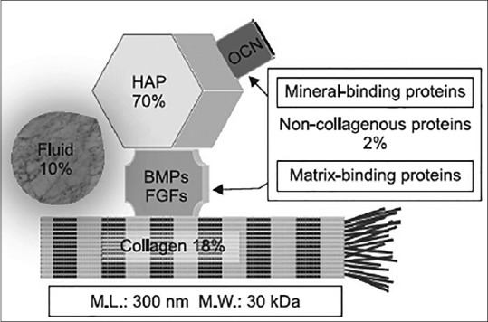
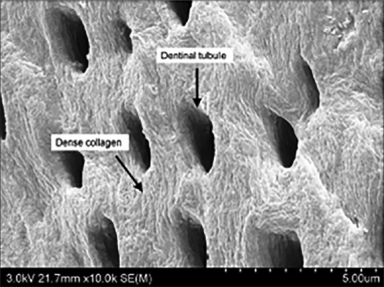
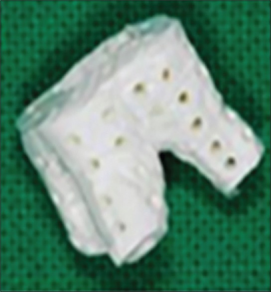
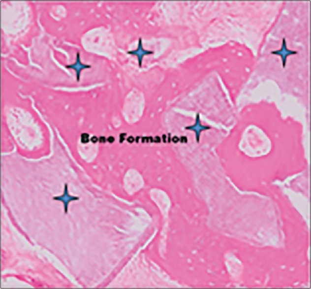
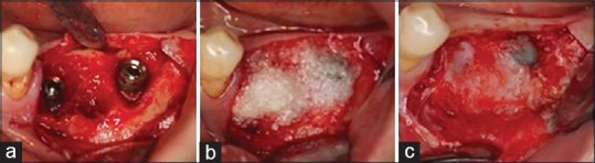
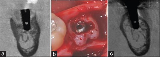
From the point of view of implant dentistry, this review discusses the development and clinical use of demineralized dentin matrix (DDM) scaffolds, produced from the patient's own extracted teeth, to repair alveolar bone defects. The structure and the organic and inorganic components of DDM are presented to emphasize the similarities with autogenous bone. Studies of DDM properties, such as osteoinductive and osteoconductive functions as well as efficacy and safety, which are mandatory for its use as a bone graft substitute, are also presented. The clinical applications of powder, block, and moldable DDM are discussed, along with future developments that can support growth factor and stem cell delivery.
Keywords: Bone graft substitute; demineralized dentin matrix; tooth.
Conflict of interest statement
There are no conflicts of interest.
Figures
References
-
- Murata M. Collagen biology for bone regenerative surgery. J Korean Assoc Oral Maxillofac Surg. 2012;38:321–5.
-
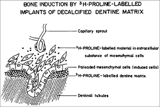
- Yeomans JD, Urist MR. Bone induction by decalcified dentine implanted into oral, osseous and muscle tissues. Arch Oral Biol. 1967;12:999–1008. - PubMed
-
- Bang G, Urist MR. Bone induction in excavation chambers in matrix of decalcified dentin. Arch Surg. 1967;94:781–9. - PubMed
-
- Urist MR, Dowell TA, Hay PH, Strates BS. Inductive substrates for bone formation. Clin Orthop Relat Res. 1968;59:59–96. - PubMed
-
- Bessho K, Tanaka N, Matsumoto J, Tagawa T, Murata M. Human dentin-matrix-derived bone morphogenetic protein. J Dent Res. 1991;70:171–5. - PubMed
Publication types
LinkOut - more resources
Full Text Sources
Other Literature Sources