

Fibroadenoma progress to ductal carcinoma in situ, infiltrating ductal carcinoma and lymph node metastasis? Report an unusual case
- PMID: 28584621
- PMCID: PMC5451661
- DOI: 10.1093/jscr/rjx064
Fibroadenoma progress to ductal carcinoma in situ, infiltrating ductal carcinoma and lymph node metastasis? Report an unusual case
Abstract
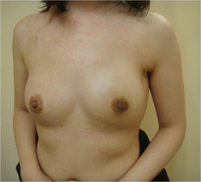
Fibroadenoma of the breast is the most common benign neoplasm in young women who present with a palpable, movable mass. Malignancy inside fibroadenomas is rare, with reported rates ranging from 0.002% to 0.125%. Carcinoma in situ inside a fibroadenoma is usually found incidentally when tumours are excised. Whether fibroadenoma is a risk factor for breast cancer remains controversial. Due to the rarity of carcinomas inside fibroadenomas, medical institutes have little experience with this phenomenon. We report an unusual case in which progression occurred from benign fibroadenoma to ductal carcinoma in situ, infiltrating ductal carcinoma and lymph node metastasis. A nipple-areolar complex-preserving mastectomy with immediate breast reconstruction with a gel implant and contralateral augmentation was performed. No local recurrence or metastasis was found during 5 years of follow-up.
Figures


References
-
- Goehring C, Morabia A. Epidemiology of benign breast disease, with special attention to histologic types. Epidemiol Rev 1997;19:310–27. - PubMed
-
- Cant PJ, Madden MV, Coleman MG, Dent DM. Non-operative management of breast masses diagnosed as fibroadenoma. Br J Surg 1995;82:792–4. - PubMed
-
- Foster ME, Garrahan N, Williams S. Fibroadenoma of the breast: a clinical and pathological study. J R Coll Surg Edinb 1988;33:16–9. - PubMed
-
- Feschenes L, Jacob S, Fabia J, Christen A. Beware of breast fibroadenomas in middle-aged women. Can J Surg 1985;28:372–4. - PubMed
-
- Dupont WD, Page DL, Parl FF, Vnencak-Jones CL, Plummer WD Jr, Rados MS, et al. . Long-term risk of breast cancer in women with fibroadenoma. N Engl J Med 1994;331:10–5. - PubMed
Publication types
LinkOut - more resources
Full Text Sources
Other Literature Sources
