

Unusual tomographic findings of complicated necrotizing pancreatitis
- PMID: 28584809
- PMCID: PMC5453663
- DOI: 10.4322/acr.2013.041
Unusual tomographic findings of complicated necrotizing pancreatitis
Abstract
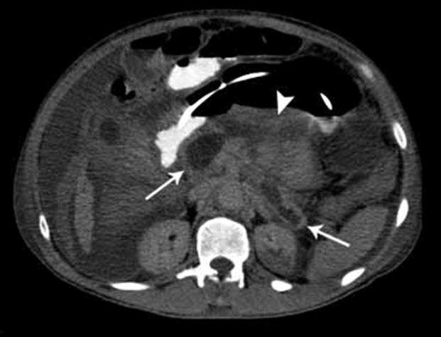
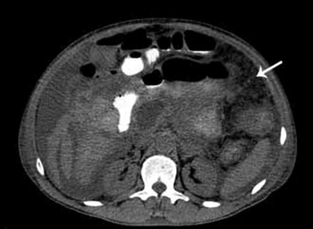
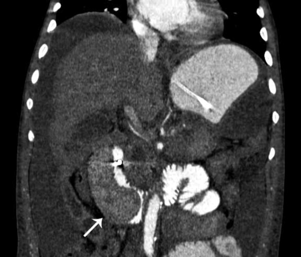
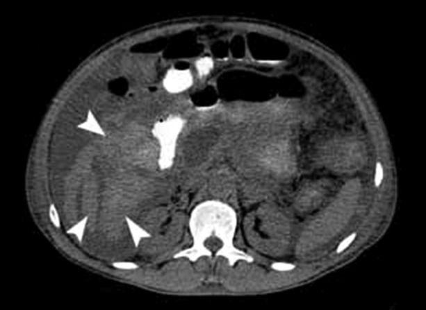
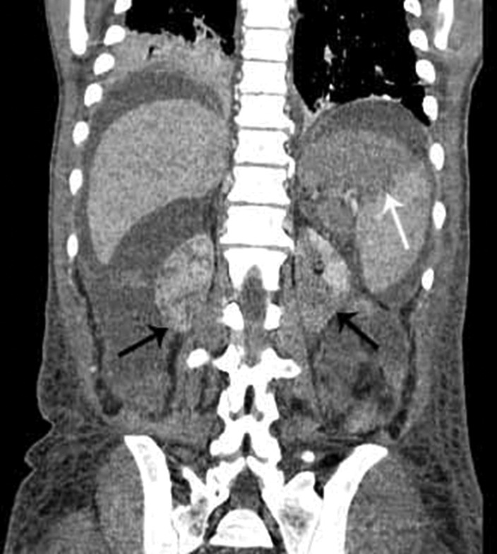
Acute pancreatitis (AP) is a potential life-threatening disease, which originates from inflammatory involvement of the pancreas and surrounding tissues. Serious complications eventuate and treatment is difficult. AP is classified in both interstitial edematous pancreatitis, which occurs in 70-80% of patients, and necrotizing pancreatitis, which occurs in 20-30% of patients. Diagnosis is based on the presence of two of the following criteria: abdominal pain, increased serum determination of amylase and/or lipase more than three times the reference value, and characteristic tomographic findings. Among the latter, there is the pancreatic and surrounding tissue damage as well as that related to distant organ involvement. This case report shows the fatal case of a male patient with a history of heavy alcoholic abuse admitted with the diagnosis of necrotizing pancreatitis. The authors call attention to the unusual tomographic findings; namely, a huge duodenal hematoma and a large hemoperitoneum, ischemic involvement of the spleen and kidneys, as well as pancreatic and peripancreatic necrosis.
Keywords: Duodenal Diseases; Hemoperitoneum; Pancreatitis, Acute Necrotizing.
Conflict of interest statement
Conflict of interest: None
Figures
Similar articles
-
Perfusion-CT--Can We Predict Acute Pancreatitis Outcome within the First 24 Hours from the Onset of Symptoms?PLoS One. 2016 Jan 19;11(1):e0146965. doi: 10.1371/journal.pone.0146965. eCollection 2016. PLoS One. 2016. PMID: 26784348 Free PMC article.
-
COVID-19 Associated Acute Necrotizing Pancreatitis with Normal Serum Amylase and Lipase Levels: Report of an Unusual Finding.Oman Med J. 2021 Sep 30;36(5):e304. doi: 10.5001/omj.2021.129. eCollection 2021 Sep. Oman Med J. 2021. PMID: 34733550 Free PMC article.
-
Involvement of thrombopoietin in acinar cell necrosis in L-arginine-induced acute pancreatitis in mice.Cytokine. 2012 Oct;60(1):294-301. doi: 10.1016/j.cyto.2012.05.005. Epub 2012 Jun 13. Cytokine. 2012. PMID: 22698803
-
2012 revision of the Atlanta classification of acute pancreatitis.Pol Arch Med Wewn. 2013;123(3):118-24. doi: 10.20452/pamw.1627. Epub 2013 Jan 25. Pol Arch Med Wewn. 2013. PMID: 23396317 Review.
-
[ACUTE PANCREATITIS IN FAMILY PHYSICIAN PRACTICE].Acta Med Croatica. 2015 Nov;69(4):357-64. Acta Med Croatica. 2015. PMID: 29083849 Review. Croatian.
References
-
- De Campos T, Parreira JG, Utiyama E, Rasslan. Pesquisa nacional sobre condutas na pancreatite aguda. Rev Col Bras Cir. 2008;35(5):304-10. Portuguese. http://dx.doi.org/10.1590/S0100-69912008000500006 - DOI
-
- Sakorafas GH, Tsiotou AG. Etiology and pathogenesis of acute pancreatitis: current concepts. J Clin Gastroenterol. 2000;30(4):343-56. - PubMed
-
- Trout AT, Elsayes KM, Ellis JH, Francis RI. Imaging of acute pancreatitis: prognostic value of computed findings. J Comput Assist Tomogr. 2010;34(4):485-95. http://dx.doi.org/10.1097/RCT.0b013e3181d344ca. - DOI - PubMed
-
- Banks PA, Freeman ML. Practice guidelines in acute pancreatitis. Am J Gastroenterol. 2006;101(10):2379-400. - PubMed
-
- Frossard JL, Steer ML, Pastor CM. Acute pancreatitis. Lancet. 2008;371(9607):143-52. - PubMed
Publication types
LinkOut - more resources
Full Text Sources
Other Literature Sources
Miscellaneous
