. 2018 Feb;34(1):98-108.
doi: 10.1007/s12264-017-0145-y. Epub 2017 Jun 5.

Sex-Dependent Glial Signaling in Pathological Pain: Distinct Roles of Spinal Microglia and Astrocytes

Affiliations

Sex-Dependent Glial Signaling in Pathological Pain: Distinct Roles of Spinal Microglia and Astrocytes

Gang Chen et al. Neurosci Bull. 2018 Feb.

Abstract

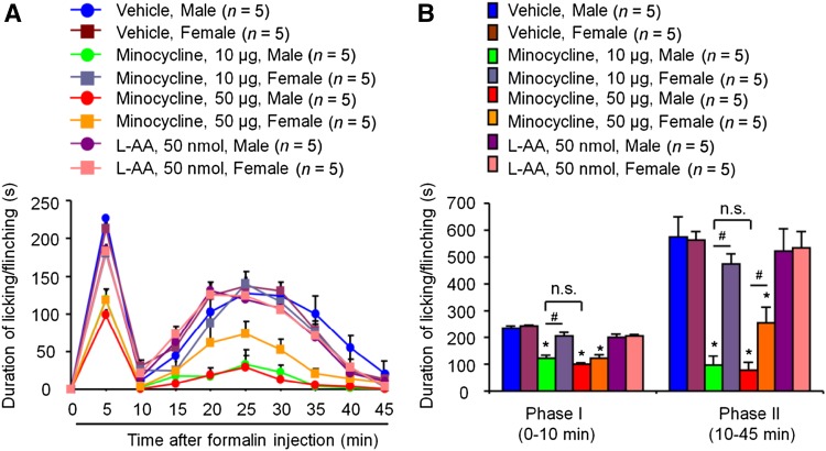
Increasing evidence suggests that spinal microglia regulate pathological pain in males. In this study, we investigated the effects of several microglial and astroglial modulators on inflammatory and neuropathic pain following intrathecal injection in male and female mice. These modulators were the microglial inhibitors minocycline and ZVEID (a caspase-6 inhibitor) and the astroglial inhibitors L-α-aminoadipate (L-AA, an astroglial toxin) and carbenoxolone (a connexin 43 inhibitor), as well as U0126 (an ERK kinase inhibitor) and D-JNKI-1 (a c-Jun N-terminal kinase inhibitor). We found that spinal administration of minocycline or ZVEID, or Caspase6 deletion, reduced formalin-induced inflammatory and nerve injury-induced neuropathic pain primarily in male mice. In contrast, intrathecal L-AA reduced neuropathic pain but not inflammatory pain in both sexes. Intrathecal U0126 and D-JNKI-1 reduced neuropathic pain in both sexes. Nerve injury caused spinal upregulation of the astroglial markers GFAP and Connexin 43 in both sexes. Collectively, our data confirmed male-dominant microglial signaling but also revealed sex-independent astroglial signaling in the spinal cord in inflammatory and neuropathic pain.

Keywords: Astrocytes; Microglia; Sex difference; Spinal cord.


Figures

Fig. 1
Fig. 1
Effects of spinal injection of microglial inhibitor and astroglial toxin on formalin-induced pain in male and female mice. A Time-course of licking and flinching behavior following intraplantar injection of 5% formalin. B Formalin-induced Phase I (1–10 min) and Phase II (10–45 min) responses. Intrathecal injection of vehicle (PBS), the microglial inhibitor minocycline (10 and 50 µg), and the astroglial toxin L-AA (50 nmol), 30 min prior to intraplantar injection of formalin. Mean ± SEM; *P < 0.05 versus corresponding control (Vehicle); # P < 0.05 vs opposite sex; n.s., not significant vs different doses; one-way ANOVA, n = 5 mice per group.
Fig. 2
Fig. 2
Effects of spinal injection of CASP6 and TNF on naïve male and female mice. A Intrathecal injection of CASP6 (5 U) and TNF (20 ng) both elicited rapid and persistent mechanical hypersensitivity in naïve males. B Intrathecal injection of TNF but not CASP6 elicited rapid and persistent mechanical hypersensitivity in naïve females. Mean ± SEM; *P < 0.05, n.s., not significant, two-way ANOVA; n = 4 mice per group; BL, baseline. Arrows indicate drug injection.
Fig. 3
Fig. 3
CASP6 contributes to formalin-induced pain and late-phase mechanical hypersensitivity in male mice. A Time course of formalin-induced pain in males and females with intrathecal vehicle or the CASP6 inhibitor ZVEID (30 µg). B ZVEID (30 µg, i.t.) reduced both formalin-induced phase I and phase II pain in males but only phase I in females. *P < 0.05, two-tailed Student’s t-test, n = 5 mice per group. C, D Formalin-induced pain was reduced in male but not female Casp6 –/– mice. Mean ± SEM; *P < 0.05, two-tailed Student’s t-test, n = 4–5 mice per group. E Formalin-induced mechanical hypersensitivity in the late phase was lower in male than in female Casp6 –/– mice. Mean ± SEM; *P < 0.05, two-way ANOVA; n = 4–5 mice per group.
Fig. 4
Fig. 4
In the CCI model, microglial inhibition reduces mechanical hypersensitivity in male but not female mice, while astroglial inhibition shows no sexual dimorphism. A CCI-induced mechanical hypersensitivity at 7 days was fully reversed by spinal injection of minocycline (50 µg = 100 nmol) in males and it was only partially reversed in females. B CCI-induced mechanical hypersensitivity at 12 days was partially reversed by L-AA (50 nmol), but there was no difference between males and females. Mean ± SEM; *P < 0.05 vs corresponding CCI 7, one-way ANOVA; # P < 0.05, n.s., not significant vs opposite sex, two-way ANOVA; n = 8 mice per group; BL, baseline. Arrows indicate drug injection.
Fig. 5
Fig. 5
Spinal inhibition of JNK, ERK, Cx43 hemichannels, and CXCR2 reduces mechanical hypersensitivity in both males and females equally in mice with chronic constriction injury. A–C CCI-induced mechanical hypersensitivity was reduced at 15 days by the JNK inhibitor D-JNKI-1 (4 nmol) (A), and at 14 days by the ERK inhibitor U0126 (25 nmol) (B); it was fully reversed at 17 days by CBX (5 µg = 8 nmol) (C), and reduced at 13 days by the CXCR2 antagonist SB225002 (20 µg = 56 nmol) (D) in males and females. E CCI-induced mechanical hypersensitivity at 14 days was not changed by vehicle (PBS) injection, and males and females did not significantly differ. Mean ±SEM; *P < 0.05 vs corresponding CCI, n.s., not significant, two-way ANOVA; n = 5–6 mice per group; BL, baseline. Arrows indicate drug injection.
Fig. 6
Fig. 6
CCI increases GFAP and Cx43 expression in the spinal dorsal horn equally in mice of both sexes. A, B Low-magnification images of double immunofluorescent staining with GFAP (red) and connexin 43 (Cx43, green) in lumbar dorsal horn sections showing both ipsilateral (Ipsi.) and contralateral (Contra.) sides in a male (A) and a female (B) 14 days after CCI (scale bars, 100 µm). A’, B’ High-magnification images (from boxes in A, B) of the double staining in the ipsilateral dorsal horn (scale bars, 50 µm). C, D Quantification of GFAP (C) and Cx43 (D) immunofluorescence from lumbar sections showing a significant increase in fluorescence intensity in the ipsilateral dorsal horn in both males and females. E Co-localization ratios of GFAP and Cx43 IR staining in the ipsilateral dorsal horn in males and females 14 days after CCI. Mean ± SEM; *P < 0.05 between ipsilateral and contralateral dorsal horn of same sex, n.s. not significant, two-tailed Student’s t-test, n = 4 mice per sex per group, 5 sections per mouse.

References

    1. Ji RR, Berta T, Nedergaard M. Glia and pain: is chronic pain a gliopathy? Pain. 2013;154(Suppl 1):S10–S28. doi: 10.1016/j.pain.2013.06.022. - DOI - PMC - PubMed
    1. Old EA, Clark AK, Malcangio M. The role of glia in the spinal cord in neuropathic and inflammatory pain. Handb Exp Pharmacol. 2015;227:145–170. doi: 10.1007/978-3-662-46450-2_8. - DOI - PubMed
    1. Ji RR, Chamessian A, Zhang YQ. Pain regulation by non-neuronal cells and inflammation. Science. 2016;354:572–577. doi: 10.1126/science.aaf8924. - DOI - PMC - PubMed
    1. Ren K, Dubner R. Interactions between the immune and nervous systems in pain. Nat Med. 2010;16:1267–1276. doi: 10.1038/nm.2234. - DOI - PMC - PubMed
    1. Ren K, Dubner R. Activity-triggered tetrapartite neuron-glial interactions following peripheral injury. Curr Opin Pharmacol. 2016;26:16–25. doi: 10.1016/j.coph.2015.09.006. - DOI - PMC - PubMed

MeSH terms

LinkOut - more resources