. 2017 Aug;16(4):859-869.
doi: 10.1111/acel.12618. Epub 2017 Jun 6.

Deletion of ghrelin prevents aging-associated obesity and muscle dysfunction without affecting longevity

Affiliations

Deletion of ghrelin prevents aging-associated obesity and muscle dysfunction without affecting longevity

Bobby Guillory et al. Aging Cell. 2017 Aug.

Abstract

During aging, decreases in energy expenditure and locomotor activity lead to body weight and fat gain. Aging is also associated with decreases in muscle strength and endurance leading to functional decline. Here, we show that lifelong deletion of ghrelin prevents development of obesity associated with aging by modulating food intake and energy expenditure. Ghrelin deletion also attenuated the decrease in phosphorylated adenosine monophosphate-activated protein kinase (pAMPK) and downstream mediators in muscle, and increased the number of type IIa (fatigue resistant, oxidative) muscle fibers, preventing the decline in muscle strength and endurance seen with aging. Longevity was not affected by ghrelin deletion. Treatment of old mice with pharmacologic doses of ghrelin increased food intake, body weight, and muscle strength in both ghrelin wild-type and knockout mice. These findings highlight the relevance of ghrelin during aging and identify a novel AMPK-dependent mechanism for ghrelin action in muscle.

Keywords: Sarcopenia; frailty; growth hormone; growth hormone secretagogue receptor; inflammation; wasting.


Figures

Figure 1
Figure 1
(A‐F). Body weight and composition in young and old ghrelin WT and KO animals. 6–9 months (young adult) and at 21–24 months of age (old) mice were fed standard nonsoy rodent chow (n = 16/group). Body weight in age‐matched animals measured in grams (A). Body composition measured by nuclear magnetic resonance (NMR), absolute lean body mass (LBM, B), and LBM percentage from body weight (C). Fat mass (D). Grip strength measured by rodent grip dynamometer (E). Treadmill activity measured in seconds where animals ran to exhaustion with increasing speed every 2 min (F). < 0.05. *young vs. old, §P < 0.05 ghrelin wild‐type (GWT) vs. knockout (GKO).
Figure 2
Figure 2
Meal pattern monitoring and calorimetry measurements. 6‐ to 9‐month‐old (young adult) and 21‐ to 24‐month‐old (old) age‐matched mice (n = 8/group) tested by Columbus Instruments Comprehensive Lab Animal Monitoring System (CLAMS). Daily food intake adjusted by body weight (A). Energy expenditure adjusted by LBM (B). Respiratory Quotient (RQ, C) and spontaneous total locomotor activity (D). < 0.05. *young vs. old, §P < 0.05 ghrelin wild‐type (GWT) vs. knockout (GKO).
Figure 3
Figure 3
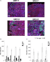
Fiber type muscle fiber distribution in young and old ghrelin WT and KO mice. Myosin heavy‐chain (MHC) immunofluorescence fiber staining (n = 4/group) on quadriceps muscle for type I fibers (blue), IIa fibers (oxidative, fatigue resistant, green), IIb fibers (glycolytic, red), and IIx fibers (black, A). Total fiber area distribution by fiber type: Ix, IIa, IIb, and IIx (μm2) (B). Total fiber number by fiber type: Ix, IIa, IIb, and IIx (C). < 0.05. *young vs. old.
Figure 4
Figure 4
AMPK, IGF‐1, and downstream mediator protein and transcript level expression in muscles from young and old ghrelin WT and KO mice. Representative Western blots probed for p‐AMPK and FAS protein levels (A). Protein densitometry quantification for pAMPK (B) and FAS (C). Transcript level expression by RT‐qPCR for pdk‐4 (D). Representative Western blots probed for p‐AKT, p‐FoxO‐1, MuRF‐1, and MafBx (atrogin) protein levels (E). Serum IGF‐1 levels by ELISA (F). Transcript level expression by RT‐qPCR for igf‐1Ea (G). Protein densitometry quantification for p‐AKT (H), p‐FoxO‐1 (I), MuRF‐1 (J), atrogin (K). Samples normalized to GAPDH (n = 4/group). < 0.05. *young vs. old, §P < 0.05 ghrelin wild‐type (GWT) vs. knockout (GKO).
Figure 5
Figure 5
Effect of acylated ghrelin administration to old ghrelin WT and KO mice. Acylated ghrelin was administered (0.8 mg kg−1 twice a day, subcutaneously) for 28 days to 21‐ to 24‐month‐old, age‐matched mice (n = 4/group). Effects of ghrelin on daily food intake (g, A), Body weight change from baseline (%, B), Lean mass change from baseline by NMR (%, C), Fat mass change from baseline by NMR (%, D), Grip strength change from baseline (%, E), Endurance change from baseline measured by treadmill (%, F). < 0.05. *young vs. old, §P < 0.05 ghrelin wild‐type (GWT) vs. knockout (GKO). C: Control, G Ghrelin‐treated.
Figure 6
Figure 6
Ghrelin deletion does not affect longevity in mice. Days survived for ghrelin WT and KO mice assessed by a Kaplan–Meier curve.

References

    1. Ballak SB, Degens H, de Haan A, Jaspers RT (2014) Aging related changes in determinants of muscle force generating capacity: a comparison of muscle aging in men and male rodents. Ageing Res. Rev. 14, 43–55. - PubMed
    1. Barazzoni R, Bosutti A, Stebel M, Cattin MR, Roder E, Visintin L, Cattin L, Biolo G, Zanetti M, Guarnieri G (2005) Ghrelin regulates mitochondrial‐lipid metabolism gene expression and tissue fat distribution favoring triglyceride deposition in liver but not skeletal muscle. Am. J. Physiol. Endocrinol. Metab. 288, E228–235. - PubMed
    1. Baumgartner RN, Wayne SJ, Waters DL, Janssen I, Gallagher D, Morley JE (2004) Sarcopenic obesity predicts instrumental activities of daily living disability in the elderly. Obes. Res. 12, 1995–2004. - PubMed
    1. Beharka AA, Meydani M, Wu D, Leka LS, Meydani A, Meydani SN (2001) Interleukin‐6 production does not increase with age. J. Gerontol. A Biol. Sci. Med. Sci. 56, B81–B88. - PubMed
    1. Bergmeister KD, Groger M, Aman M, Willensdorfer A, Manzano‐Szalai K, Salminger S, Aszmann OC (2016) Automated muscle fiber type population analysis with ImageJ of whole rat muscles using rapid myosin heavy chain immunohistochemistry. Muscle Nerve 54, 292–299. - PubMed

Publication types

MeSH terms

LinkOut - more resources