. 2017 Jul;6(7):1583-1594.
doi: 10.1002/sctm.16-0449. Epub 2017 Jun 6.

Liver-Directed Human Amniotic Epithelial Cell Transplantation Improves Systemic Disease Phenotype in Hurler Syndrome Mouse Model

Affiliations

Liver-Directed Human Amniotic Epithelial Cell Transplantation Improves Systemic Disease Phenotype in Hurler Syndrome Mouse Model

Natalie S Rodriguez et al. Stem Cells Transl Med. 2017 Jul.

Abstract

Mucopolysaccharidosis type 1 (MPS1) is an inherited lysosomal storage disorder caused by a deficiency in the glycosaminoglycan (GAG)-degrading enzyme α-l-iduronidase (IDUA). In affected patients, the systemic accumulation of GAGs results in skeletal dysplasia, neurological degeneration, multiple organ dysfunction, and early death. Current therapies, including enzyme replacement and bone marrow transplant, improve life expectancy but the benefits to skeletal and neurological phenotypes are limited. In this study, we tested the therapeutic efficacy of liver-directed transplantation of a placental stem cell, which possesses multilineage differentiation potential, low immunogenicity, and high lysosomal enzyme activity. Unfractionated human amniotic epithelial cells (hAECs) were transplanted directly into the liver of immunodeficient Idua knockout mouse neonates. The hAECs engraftment was immunohistochemically confirmed with anti-human mitochondria staining. Enzyme activity assays indicated that hAECs transplantation restored IDUA function in the liver and significantly decreased urinary GAG excretion. Histochemical and micro-computed tomography analyses revealed reduced GAG deposition in the phalanges joints and composition/morphology improvement of cranial and facial bones. Neurological assessment in the hAEC treated mice showed significant improvement of sensorimotor coordination in the hAEC treated mice compared to untreated mice. Results confirm that partial liver cell replacement with placental stem cells can provide long-term (>20 weeks) and systemic restoration of enzyme function, and lead to significant phenotypic improvement in the MPS1 mouse model. This preclinical data indicate that liver-directed placental stem cell transplantation may improve skeletal and neurological phenotypes of MPS1 patients. Stem Cells Translational Medicine 2017;6:1583-1594.

Keywords: Amnion; Cell transplantation; Congenital; Metabolism; Mucopolysaccharidosis I; Placenta.


Figures

Figure 1
Figure 1
Primary hAEC characterization and the liver‐directed cell transplantation. (A): Single cell gene expression data reveal IDUA gene expressions in hAECs is equivalent to that of primary human hepatocytes. Delta‐Ct values were calculated from the difference in the Ct of IDUA and that of three housekeeping genes: GAPDH, beta‐actin, and cyclophilin A. Each dot represents the delta‐Ct value of a single cell that was isolated from three different placentae. Three different primary human hepatocytes purchased from Gibco/Thermo Fisher Scientific were used as the control. (B): Western blot analysis demonstrated that hAECs contain more IDUA protein than hepatocytes. (C): In vitro IDUA enzyme activity is not significantly different in hAEC and primary hepatocytes. Mean  ± SEM were graphed. (ns p =.9543, Unpaired Student's t test). (D): The schematic shows an overview of the experimental procedure. The amniotic epithelial cells were enzymatically isolated from the innermost lining of placenta (H&E staining). The isolated cells were cryopreserved immediately and thawed 48–72 hours before cell transplantation. A total of one million hAECs were transplanted twice by direct percutaneous injection into the neonate mouse liver parenchyma at days 2 and 5 after birth. Mice were observed until 28 weeks after the transplantation. At 28 weeks after liver‐directed injection, NOD/SCID MPS1 mice were euthanized, livers were processed for histology, and engrafted human cells were detected using a mouse anti‐human mitochondrial antibody with NovaRED peroxidase substrate. The panel shows PBS injected control mouse liver (E) and hAEC injected mouse liver (F). Abbreviations: CV, central vein; GAPDH, glyceraldehyde‐3‐phosphate dehydrogenase; hAECs, human amniotic epithelial cells; IDUA, α‐l‐iduronidase; PV, portal vein.
Figure 2
Figure 2
Restoration of IDUA enzyme function in MPS1 homozygous mice after human amniotic epithelial cells (hAECs) transplantation. (A): hAEC transplanted MPS1 homozygous (Treated) mice demonstrated significant decrease of urinary GAG concentration. (B): Throughout the monitoring period of 8 to 24 weeks, urinary GAG concentrations of treated mice were consistently lower than untreated mice. The average urinary GAG concentrations of untreated mice (3.112  ± 1.850 μg GAG/mg creatinine) and wild type (0.531  ± 0.357 μg GAG/mg creatinine) were supplementally presented in red and blue straight lines, respectively. (C): Tissue IDUA activity was biochemically assayed in six major organs including liver, kidney, lung, spleen, heart, and brain. While IDUA enzyme activities were higher in all organs of treated mice, the differences were not statistically significant except in the liver. Mean ± SEM were graphed (*, p <.05, Unpaired Student's t test). (D): Although liver IDUA enzyme activity was significantly restored by hAEC transplantation, it was still significantly lower than the wild type mouse liver. Mean ± SEM were graphed. n = 26. (E): Liver histology (Alcian Blue staining of mucopolysaccharides and nuclear fast red counterstain) did not demonstrate notable decreases in GAG accumulation (indicated by yellow arrows). Abbreviations: BD, bile duct; CV, central vein; GAG, glycosaminoglycan; IDUA, α‐L‐iduronidase; PV, portal vein; WT, wild type.
Figure 3
Figure 3
Bone morphology and quality analysis by high‐resolution X‐ray micro‐computed tomography (micro‐CT). (A): Increased zygomatic bone volume is a signature phenotype of homozygous Idua knockout (MSP1) mice. We measured both right (blue) and left (green) zygomatic bone volume and density using micro‐CT derived virtual models. (B): Zygomatic bone volume was significantly decreased in treated mice. Volume measured as the product of the number of voxels making the zygomatic bone and the voxel resolution. Each dot represents zygomatic bone volume normalized with skull size. n = 26. (C): Sagittal view bone surface renderings reveal the short, flattened nose phenotype in untreated and to a lesser extent, treated mice. Comparative analysis of the volumetric bone density distribution reveal higher bone density level in the untreated mouse skull compared to WT and treated mouse. The results were presented using a common colormap across the untreated WT and treated mice that rendered all bone densities between low bone density value (–1,000 raw CT units as red) to high bone density values (8,000 raw CT units as yellow) volume renderings of bone density. Note the increase in the amount of yellow in the untreated compared to WT and treated mouse. (D): HA density per a cubic centimeter indicated statistically significant differences between treated and untreated groups. Mineral density was calibrated in units of mg HA/cc using a standard hydroxyapatite phantom (Scanco Medical AG). Mean ± SEM were graphed, n = 26. Abbreviations: HA, hydroxyapatite; MPS1, mucopolysaccharidosis type 1; WT, wild type.
Figure 4
Figure 4
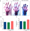
Limb skeletal morphology in MPS1 mice demonstrated by Alizarin Red and Alcian Blue staining. (A): Forelimbs of WT; MPS1 homozygous treated (Treated); and MPS1 Homozygous (Untreated) mice were stained with Alizarin Red (bone) and Alcian Blue (acid mucopolysaccharides). Arthrigryposis in the first and second joints of the forelimb with “claw‐shaped” hand as well as thicker connective tissue deposition were seen in the MPS‐1 homozygous untreated mice. The width of the third metacarpal (B) and proximal phalanges (C) were not statistically significant different between groups, though slight improvement was seen in treated mice versus untreated mice (Mean ± SEM, n = 14 per group, p  =.1021). Abbreviations: MPS1, mucopolysaccharidosis type 1; WT, wild type.
Figure 5
Figure 5
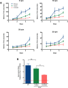
Rotarod performance test showing behavioral phenotype improvement in the human amniotic epithelial cells (hAECs) transplanted mice. (A); Wild type (WT; Blue), mucopolysaccharidosis type 1 (MPS1) homozygous treated (Treated; Green) and MPS1 homozygous (Untreated; Red) were trained on the rotarod for 5 days at 4 different rotation speeds (5, 15, 25, 35 rpm). All mice were 23 weeks of age and naive to the rotarod test. With all tested speeds on the rotarod, the treated mice showed improvement in motor coordination skills. (B): After 5 days of a learning period, the improvement ratio was significantly higher in the hAEC‐transplanted mice (green) compared to untreated (red) mice at a speed of 15 rpm. The improvement was demonstrated as relative to Day 1 balance time. There was no significant difference between wild type mice and treated mice. Mean  ± SEM were graphed (ns p  .05, **, p < .01, one‐way ANOVA).

References

    1. Dorfman A, Matalon R. The mucopolysaccharidoses (a review). Proc Natl Acad Sci USA 1976;73:630–637. - PMC - PubMed
    1. Bie H, Yin J, He X et al. Insights into mucopolysaccharidosis I from the structure and action of α‐L‐iduronidase. Nat Chem Biol 2013;9:739–745. - PMC - PubMed
    1. Meikle PJ, Hopwood JJ, Clague AE et al. Prevalence of lysosomal storage disorders. JAMA 1999;281:249–254. - PubMed
    1. Wraith JE, Jones S. Mucopolysaccharidosis type I. Pediatr Endocrinol Rev 2014;12(suppl 1):102–106. - PubMed
    1. Pastores GM. Laronidase (Aldurazyme): Enzyme replacement therapy for mucopolysaccharidosis type I. Expert Opin Biol Ther 2008;8:1003–1009. - PubMed

Publication types

LinkOut - more resources