. 2017 Dec;16(6):987-996.
doi: 10.1177/1533034617712397. Epub 2017 Jun 6.

Electrosensitization Increases Antitumor Effectiveness of Nanosecond Pulsed Electric Fields In Vivo

Affiliations

Electrosensitization Increases Antitumor Effectiveness of Nanosecond Pulsed Electric Fields In Vivo

Claudia Muratori et al. Technol Cancer Res Treat. 2017 Dec.

Abstract

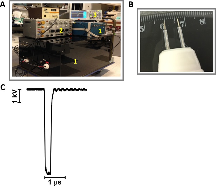
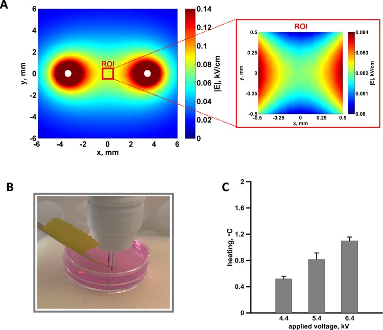
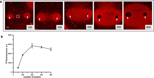
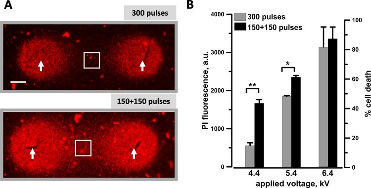
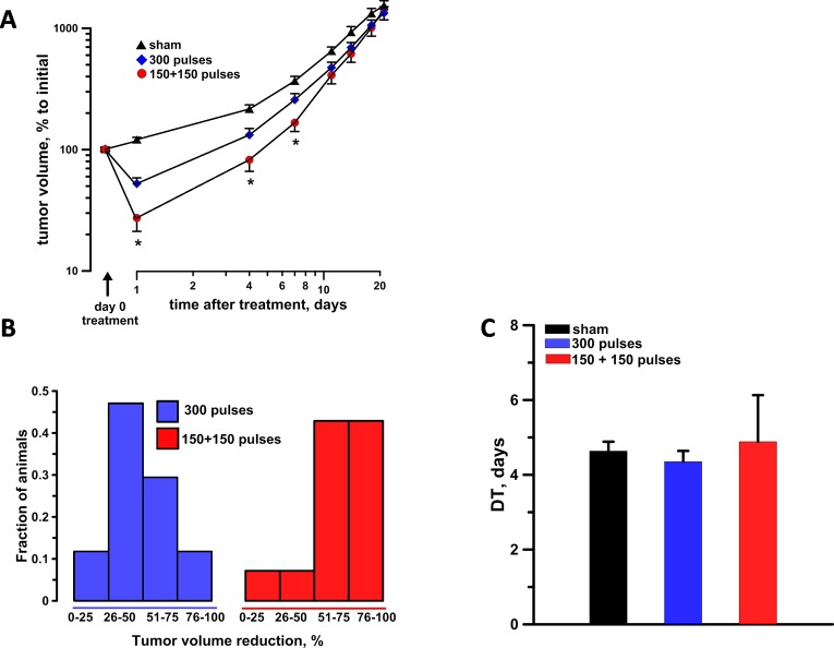
Nanosecond pulsed electric fields are emerging as a new modality for tissue and tumor ablation. We previously reported that cells exposed to pulsed electric fields develop hypersensitivity to subsequent pulsed electric field applications. This phenomenon, named electrosensitization, is evoked by splitting the pulsed electric field treatment in fractions (split-dose treatments) and causes in vitro a 2- to 3-fold increase in cytotoxicity. The aim of this study was to show the benefit of split-dose treatments for in vivo tumor ablation by nanosecond pulsed electric field. KLN 205 squamous carcinoma cells were embedded in an agarose gel or grown subcutaneously as tumors in mice. Nanosecond pulsed electric field ablations were produced using a 2-needle probe with a 6.5-mm interelectrode distance. In agarose gel, splitting a pulsed electric field dose of 300, 300-ns pulses (20 Hz, 4.4-6.4 kV) in 2 equal fractions increased cell death up to 3-fold compared to single-train treatments. We then compared the antitumor effectiveness of these treatments in vivo. At 24 hours after treatment, sensitizing tumors by a split-dose pulsed electric field exposure (150 + 150, 300-ns pulses, 20 Hz, 6.4 kV) caused a 4- and 2-fold tumor volume reduction as compared to sham and single-train treatments, respectively. Tumor volume reduction that exceeds 75% was 43% for split-dose-treated animals compared to only 12% for single-dose treatments. The difference between the 2 experimental groups remained statistically significant for at least 1 week after the treatment. The results show that electrosensitization occurs in vivo and can be exploited to assist in vivo cancer ablation.

Keywords: electrosensitization; irreversible electroporation; nanoporation; nanosecond pulsed electric fields (nsPEF); tumor ablation.


Conflict of interest statement

Declaration of Conflicting Interests: The author(s) declared no potential conflicts of interest with respect to the research, authorship, and/or publication of this article.

Figures

Figure 1.
Figure 1.
Pulsed electric field (PEF) exposure system. A, The pulse generator (1) was triggered externally from a stimulator (2), and the pulse amplitude and shape were monitored using a digital oscilloscope (3). B, The 2-needle probe showing the 6.5 mm separation. C, The shape of the electric pulse at 6.4 kV.
Figure 2.
Figure 2.
The electric field distribution (A) and temperature measurements (B and C) in 3-dimensional (3D) cell cultures. The electric field distribution in the plane perpendicular to the needle electrodes (white circles) and 3.8 mm above the bottom of the Petri dish (left) and within the 1 mm2 region of interest (ROI) used for the quantification of the propidium (Pr) fluorescence (right). Note that the 2 maps have different color scales. For 100 V applied between the electrodes, the mean electric field in the ROI was 0.082 kV/cm with 1.2% variation. B, The fiber optic probe, used to measure the temperature, placed in the center of the gap between the electrodes. C, Temperature rise measured after 300, 300 ns pulses (20 Hz) delivered at the indicated voltages.
Figure 3.
Figure 3.
Analysis of the cytotoxic effect of 300 ns, 6.4 kV pulses in KLN 205 cells embedded in agarose. A, The ablation area between and around the 6.5-mm gap pulsed electric field (PEF)–delivering electrodes (arrows) was visualized by propidium (Pr) uptake by dead cells. Images were taken 2 hours after exposure to the indicated number of pulses. Scale bar: 1 mm. B, Quantification of the cytotoxic effect by the mean intensity of Pr fluorescence (as measured within the region of interest shown in (A). Mean ± standard error (SE), n = 3. Note signal saturation at 400 to 800 pulses, which indicates killing of 100% of cells within the studied region.
Figure 4.
Figure 4.
Electrosensitization efficiency at different applied voltages when using a pulsed electric field (PEF)–delivering probe with 6.5 mm gap between the electrodes. KLN 205 cells seeded in 1% agarose were exposed to either a single train of 300 pulses (300 ns, 20 Hz) or 2 trains of 150 pulses each with a 2-minute interval. The pulse amplitude was varied from 4.4 to 6.4 kV. A, Each sample at 4.4 kV, a representative propidium (Pr) fluorescence image. The arrows identify the nanosecond pulsed electric field (nsPEF) delivering electrodes. Scale bar: 1 mm. The quantification in (B) shows the Pr uptake (left Y-axis) and the percentage of cell death (right Y-axis) measured within the region of interest (white square) shown in (A). Mean ± standard error (SE), n = 5 to 6. *P < .01 and **P < .001.
Figure 5.
Figure 5.
In vivo split-dose treatments engaging electrosensitization increase the antitumor effectiveness of nsPEF. A, Tumor growth curves in sham exposure, single-dose 300 pulses (300 ns, 6.4 kV, 20 Hz), and split-dose 150 + 150 pulses with 2-minute interval experimental groups. For each animal, the data were normalized to the tumor volume measured immediately before treatment. B, The tumor reduction frequency histogram at 24 hours after the treatment. C, The tumor doubling time. Mean ± standard error (SE), n = 15, 17, and 14 for sham, split- and single-dose groups, respectively. *P < .05 for the difference between single- and split-dose groups.

References

    1. Knavel EM, Brace CL. Tumor ablation: common modalities and general practices. Tech Vasc Interv Radiol. 2013;16(4):192–200. - PMC - PubMed
    1. Jiang C, Davalos RV, Bischof JC. A review of basic to clinical studies of irreversible electroporation therapy. IEEE Trans Biomed Eng. 2015;62(1):4–20. - PubMed
    1. Rubinsky B. Irreversible electroporation in medicine. Technol Cancer Res Treat. 2007;6(4):255–260. - PubMed
    1. Bhonsle S, Bonakdar M, Neal RE, II, et al. Characterization of irreversible electroporation ablation with a validated perfused organ model. J Vasc Interv Radiol. 2016;27(12):1913–1922.e2. - PubMed
    1. Kos B, Voigt P, Miklavcic D, Moche M. Careful treatment planning enables safe ablation of liver tumors adjacent to major blood vessels by percutaneous irreversible electroporation (IRE). Radiol Oncol. 2015;49(3):234–241. - PMC - PubMed