. 2017 Jul;27(7):933-945.
doi: 10.1038/cr.2017.81. Epub 2017 Jun 6.

One-step generation of complete gene knockout mice and monkeys by CRISPR/Cas9-mediated gene editing with multiple sgRNAs

Affiliations

One-step generation of complete gene knockout mice and monkeys by CRISPR/Cas9-mediated gene editing with multiple sgRNAs

Erwei Zuo et al. Cell Res. 2017 Jul.

Abstract

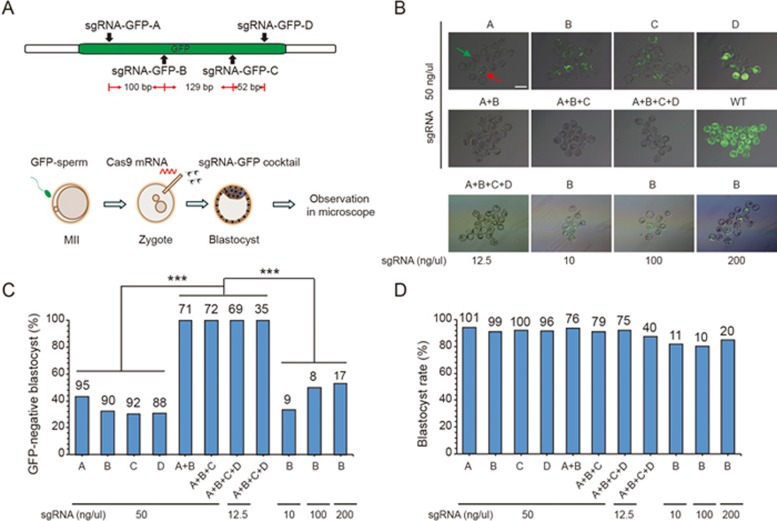
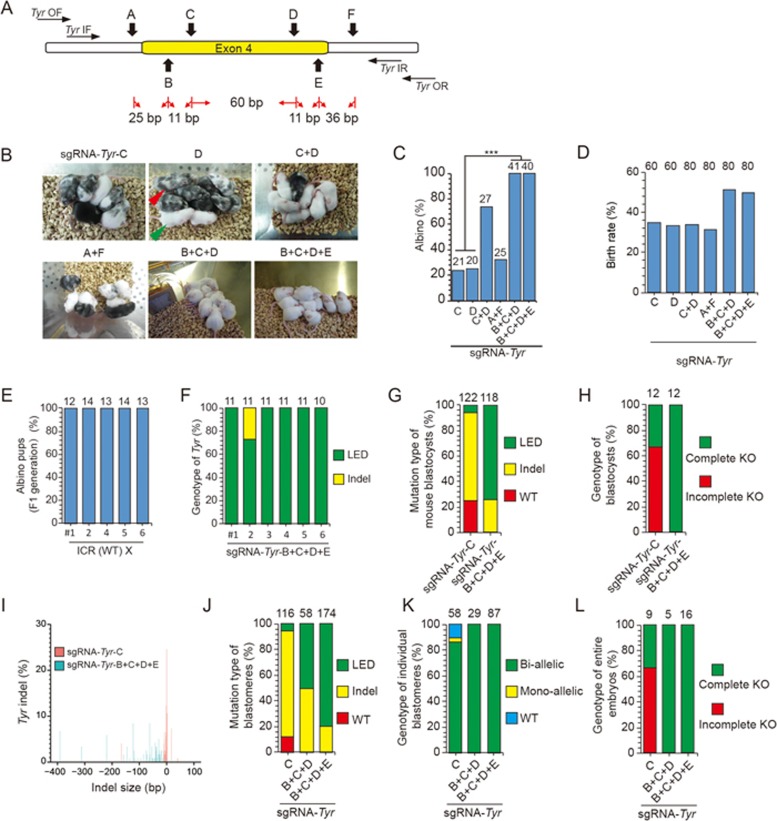
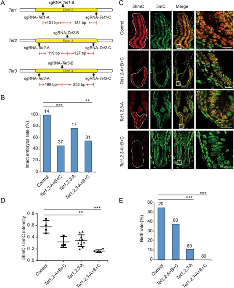
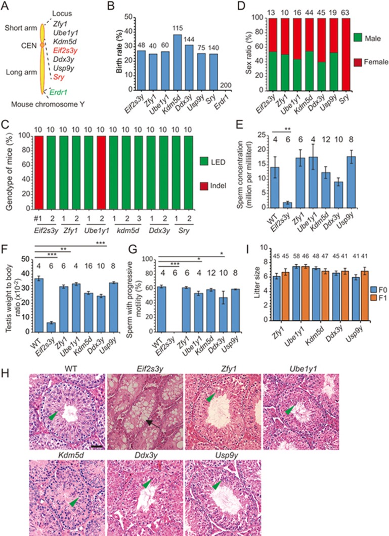
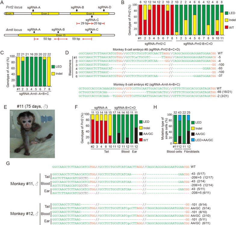
The CRISPR/Cas9 system is an efficient gene-editing method, but the majority of gene-edited animals showed mosaicism, with editing occurring only in a portion of cells. Here we show that single gene or multiple genes can be completely knocked out in mouse and monkey embryos by zygotic injection of Cas9 mRNA and multiple adjacent single-guide RNAs (spaced 10-200 bp apart) that target only a single key exon of each gene. Phenotypic analysis of F0 mice following targeted deletion of eight genes on the Y chromosome individually demonstrated the robustness of this approach in generating knockout mice. Importantly, this approach delivers complete gene knockout at high efficiencies (100% on Arntl and 91% on Prrt2) in monkey embryos. Finally, we could generate a complete Prrt2 knockout monkey in a single step, demonstrating the usefulness of this approach in rapidly establishing gene-edited monkey models.


Figures

Figure 1
Figure 1
Complete deletion of GFP in GFP-embryos by C-CRISPR. (A) Schematic diagram of sgRNA-targeting sites at GFP locus and experimental design. Cas9 mRNA and single or multiple sgRNAs-targeting GFP were injected into individual mouse zygotes and GFP signal was examined at blastocyst stage. (B) Images of GFP signal of blastocysts resulting from different forms of GFP targeting. Examples of GFP-negative blastocyst (green arrow, no GFP signal in any cells of the blastocyst) and GFP-positive blastocyst with mosaicism (red arrow, GFP signal in some cells of the blastocyst) are shown in the first image. The concentration of each sgRNA was shown in each group. Scale bar, 200 μm. (C) Histograms showing percentages of GFP-negative blastocysts (without mosaicism) resulting from different forms of GFP targeting. Two or more targeting sgRNAs (sgRNA-GFP-A+B, A+B+C, or A+B+C+D) resulted in higher percentages of GFP-negative blastocysts than single sgRNA targeting (sgRNA-GFP-A, B, C, or D). Number above the bar, total number of blastocysts counted (***P < 0.001, χ2 test). The concentration of each sgRNA was shown at the bottom. (D) Blastocyst rate of embryos resulting from different GFP targeting. Number, total number of zygotes injected. The concentration of each sgRNA was shown at the bottom.
Figure 2
Figure 2
One-step generation of Tyr complete knockout mice by C-CRISPR. (A) Schematic diagram of sgRNA-targeting sites at Tyr locus. The space between each sgRNA is from the end of last base of one sgRNA to the first base of another sgRNA (including PAM sequence). (B) Representative results of pigmentation phenotypes of mice resulting from Tyr targeting. Green arrowhead, albino; red arrowhead, mosaic pigmentation. (C) Histograms showing the percentage of albino mice resulting from Tyr targeting. Two or more targeting sgRNAs (sgRNA-Tyr-C+D, B+C+D, or B+C+D+E) resulted in higher percentage of albino mice than single-sgRNA targeting (sgRNA-Tyr-C or D). Number, total number of mice counted (***P < 0.001, χ2 test). (D) Birth rate of mice resulting from Tyr targeting. Number, total number of embryos transferred. (E) Reproductive ability and germline transmission of Tyr knockout mice by C-CRISPR. Five gene-modified mice (sgRNA-Tyr-B+C+D+E) were mated with WT ICR mice individually. Each pair of mice gave birth to two litters of pups. (F) Albino mice resulting from sgRNA-Tyr-B+C+D+E targeting were sequenced. DNA was isolated from tails of albino mice and PCR amplified. #2 mice contained large-fragment exon deletion and indels. LED, large-fragment exon deletion; Indels, insertion, or deletion of bases. Number above each column, total TA clones sequenced. (G and H) Percentages of different mutation types (G) and genotypes (H) of blastocysts resulting from Tyr targeting. Number, total TA clones sequenced and analyzed. WT, wild-type allele. Complete KO, blastocyst with complete knockout mutations; Incomplete KO, blastocyst with WT allele and knockout mutations. Number, total blastocysts counted. (I) Size distribution of deletions or insertions in blastocysts with Tyr targeting. (J and K) Percentages of different mutation types (J) and genotypes (K) for individual blastomeres of 8- to 16-cell embryos with Tyr targeting. LED, large-fragment exon deletion; Bi-allelic, bi-allelic mutations; Mono-allelic, mono-allelic mutations. Number, total alleles, or blastomeres analyzed. (L) Percentages of different genotypes of entire 8- to 16-cell embryos with Tyr targeting. Number, total embryos analyzed.
Figure 3
Figure 3
One-step generation of mice with complete triple knockout of Tet genes by C-CRISPR. (A) Schematic diagram of sgRNA-targeting sites at Tet1, Tet2 and Tet3 loci, with three sgRNAs for each locus. (B) Percentages of control and Tet gene-edited E7.5 embryos that were intact. Number, total number of embryos counted (***P < 0.001, **P < 0.01, χ2 test). (C) Immunostaining of tissue sections showing the level of 5hmC (red) and 5mC (green) in E7.5 embryos with different forms of Tet gene targeting. White-dashed line, epiblast of E7.5 embryos, with boxed regions shown at a higher resolution on the right. “5hmC”, 5-hydroxymethylcytosine; “5mC”, 5-methylcytosine; scale bar, 50 μm. (D) The relative levels of 5mC and 5hmC in E7.5 embryos. Each data point represents the ratio (5hmC/5mC) of the average immunofluorescence intensities measured from one tissue section. Error bars, SEM (***P < 0.001, **P < 0.01, unpaired t-test). (E) Birth rate of mice for control embryos and different forms of Tet gene targeting. Number: total number of embryos transferred to surrogates (***P < 0.001, χ2 test).
Figure 4
Figure 4
Phenotype analysis of F0 mice with individual Y chromosome gene deletion by C-CRISPR. (A) Schematic diagram of eight targeted genes in Y chromosome. Two sgRNAs were designed to target each gene. Red, gene previously studied with conventional knockout mice; green, gene located in both X and Y chromosomes. (B) Birth rate of mice with embryos edited by the C-CRISPR method for deletion of individual Y-chromosome gene. Number, total embryos transferred. Erdr1 targeting resulted in embryonic lethality. (C) Sex ratio of mice with deletion of different Y-chromosome genes. (D) DNA sequence analysis of gene-edited mice in Y chromosome by C-CRISPR. Tails of mice resulting from different gene targeting in Y chromosome were genotyped. LED, large-fragment exon deletion; Indels, insertion, or deletion of bases. Number above each column, total TA clones sequenced. (E-G) Sperm concentration (E) weight-to-body ratio of testis (F) and percentages of sperm with progressive motility (G) for mice with deletion of different Y-chromosome genes. Number, total number of samples counted (***P < 0.001, **P < 0.01, *P < 0.05, χ2 test). (H) Histological analysis of the testis sections from adult mice with deletion of different Y-chromosome genes. Arrow, abnormal vacuoles of seminiferous tubule; arrowhead, sperm. Abnormality was found only in the Eif2s3y deleted testis. Bar, 20 μm. (I) Reproductivity of F0 and F1 mice with deletion of different Y-chromosome genes. Number, total pups obtained.
Figure 5
Figure 5
One-step generation of complete knockout monkey by C-CRISPR. (A) Schematic diagram of sgRNA-targeting sites at Prrt2 and Arntl loci in monkey. (B and C) Percentages of different mutation types for 8-cell embryos with Prrt2 (B) and Arntl (C) targeting. LED, large-fragment exon deletion. Indels, insertion, or deletion of bases. Number above each column, total alleles analyzed from blastomeres of 8-cell embryos with Prrt2 targeting or total TA clones sequenced from 8-cell embryos with Arntl targeting. (D) Genotyping analysis on single blastomeres of 8-cell embryos targeted with sgRNA-Prrt2-B+C+D or whole 8-cell embryos targeted with sgRNA-Arntl-A+B+C. The sgRNA targeted sequences are labeled in green and the PAM sequences are labeled in red; deleted nucleotides are indicated by hyphens. Dashed lines mark the region omitted for clarity. (E) Photograph of Prrt2-knockout monkey (#11). (F) Percentages of different mutation types found in tail, ear, and blood cells from aborted and live monkeys by Prrt2 targeting. Aborted monkeys with sgRNA-Prrt2-A targeting: monkey #2, #3; live monkeys with sgRNA-Prrt2-A targeting: monkey #4, #8, #10; live monkeys with sgRNA-Prrt2-B+C+D targeting: monkey #11, #12. AA/GC, a point mutation in Prrt2(335N to 335A). (G) Representative sequences from ear, tail, and blood cells of monkey #11 and #12 with sgRNA-Prrt2-B+C+D targeting. The sgRNA-targeting sequences are labeled in green and PAM sequences are labeled in red; deleted nucleotides are indicated by hyphens. Dashed lines mark the region omitted for clarity. (H) Single-cell analysis on blood cells and fibroblasts from two live monkeys with Prrt2 editing (#11, #12). Number, total number of cells analyzed.

References

    1. Wang H, Yang H, Shivalila CS, et al. One-step generation of mice carrying mutations in multiple genes by CRISPR/Cas-mediated genome engineering. Cell 2013; 153:910–918. - PMC - PubMed
    1. Niu Y, Shen B, Cui Y, et al. Generation of gene-modified cynomolgus monkey via Cas9/RNA-mediated gene targeting in one-cell embryos. Cell 2014; 156:836–843. - PubMed
    1. Yen ST, Zhang M, Deng JM, et al. Somatic mosaicism and allele complexity induced by CRISPR/Cas9 RNA injections in mouse zygotes. Dev Biol 2014; 393:3–9. - PMC - PubMed
    1. Izpisua Belmonte JC, Callaway EM, Caddick SJ, et al. Brains, genes, and primates. Neuron 2015; 86:617–631. - PMC - PubMed
    1. Jennings CG, Landman R, Zhou Y, et al. Opportunities and challenges in modeling human brain disorders in transgenic primates. Nat Neurosci 2016; 19:1123–1130. - PubMed

MeSH terms