

Brain Tissue Pulsatility is Increased in Midlife Depression: a Comparative Study Using Ultrasound Tissue Pulsatility Imaging
- PMID: 28585568
- PMCID: PMC5686485
- DOI: 10.1038/npp.2017.113
Brain Tissue Pulsatility is Increased in Midlife Depression: a Comparative Study Using Ultrasound Tissue Pulsatility Imaging
Abstract
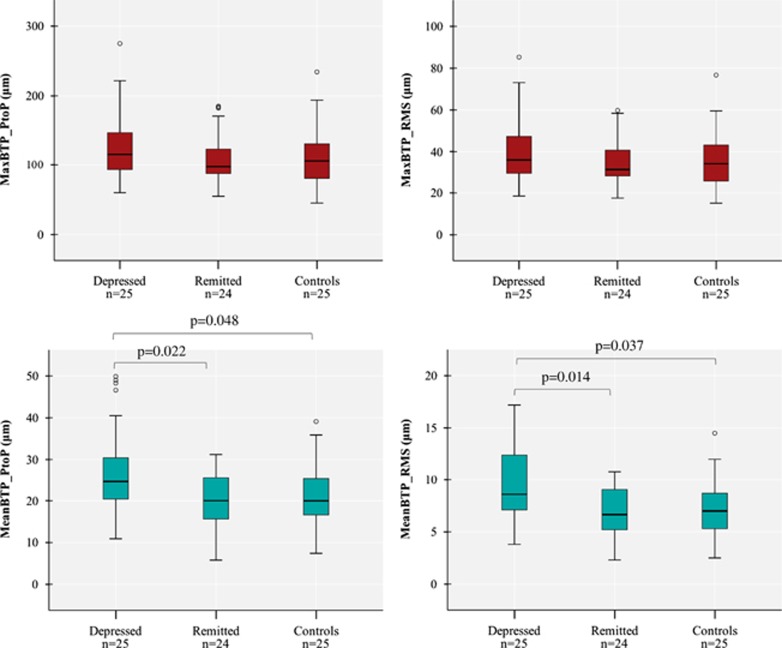
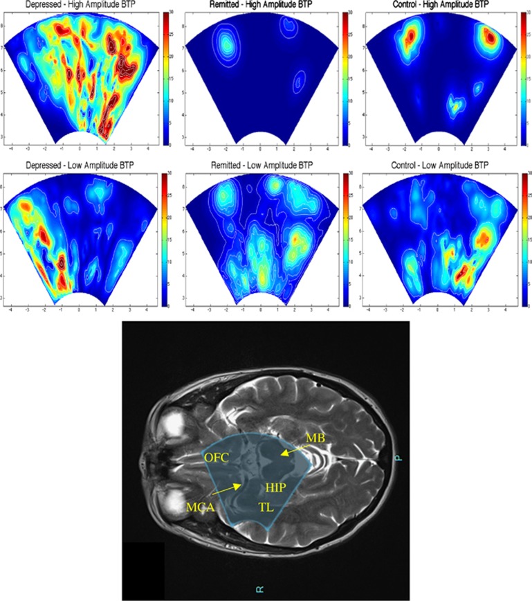
Cerebrovascular disease (CVD) is consistently associated with late-life depression but poorly documented in midlife depression. It can be hypothesized that the relatively low sensitivity of conventional neuroimaging techniques does not allow the detection of subtle CVD in midlife depression. We used tissue pulsatility imaging (TPI), a novel ultrasound (US) neuroimaging technique that has demonstrated good sensitivity to detect changes in the pulsatility of small brain volumes, to identify early and subtle changes in brain vascular function in midlife depression. We compared the maximum and mean brain tissue pulsatility (MaxBTP and MeanBTP), as identified by TPI, between three groups of middle-aged females matched for age: patients with depression (n=25), patients with remitted depression (n=24) and community controls (n=25). MRI arterial spin labeling, white matter hyperintensities (WMHs) and transcranial doppler (TCD) were used as control conventional markers for CVD. We found no difference in the MRI and TCD measures among the three groups. In contrast, depressive patients showed an increased BTP related to the mean global brain pulsatility (MeanBTP) and no change related to large vessels (MaxBTP) in comparison with the remitted and control groups. US neuroimaging is a highly accurate method to detect brain pulsatility changes related to cerebrovascular functioning, and TPI identified an increased BTP in midlife depressed patients, suggesting early and subtle vascular impairments in this population at risk for CVD such as stroke or WMHs. Because high pulsatility could represent prodromal cerebrovascular changes that damage the brain over time, this paper provides a potential target for blocking the progression of CVD.
Figures
Similar articles
-
Ultrasound Measures of Brain Pulsatility Correlate with Subcortical Brain Volumes in Healthy Young Adults.Ultrasound Med Biol. 2018 Nov;44(11):2307-2313. doi: 10.1016/j.ultrasmedbio.2018.06.016. Epub 2018 Aug 18. Ultrasound Med Biol. 2018. PMID: 30131259
-
Cerebral blood flow velocity positively correlates with brain volumes in long-term remitted depression.Prog Neuropsychopharmacol Biol Psychiatry. 2018 Feb 2;81:243-249. doi: 10.1016/j.pnpbp.2017.09.018. Epub 2017 Sep 20. Prog Neuropsychopharmacol Biol Psychiatry. 2018. PMID: 28939189 Clinical Trial.
-
Ultrasound Tissue Pulsatility Imaging Suggests Impairment in Global Brain Pulsatility and Small Vessels in Elderly Patients with Orthostatic Hypotension.J Stroke Cerebrovasc Dis. 2017 Feb;26(2):246-251. doi: 10.1016/j.jstrokecerebrovasdis.2016.09.002. Epub 2016 Nov 25. J Stroke Cerebrovasc Dis. 2017. PMID: 27894887
-
Brain structure and function and the outcomes of treatment for depression.J Clin Psychiatry. 1997;58 Suppl 16:22-31. J Clin Psychiatry. 1997. PMID: 9430506 Review.
-
Impairment of GABAergic transmission in depression: new insights from neuroimaging studies.Crit Rev Neurobiol. 2000;14(1):23-45. doi: 10.1615/critrevneurobiol.v14.i1.20. Crit Rev Neurobiol. 2000. PMID: 11253954 Review.
Cited by
-
The Effects of Hypocapnia on Brain Tissue Pulsations.Brain Sci. 2020 Sep 6;10(9):614. doi: 10.3390/brainsci10090614. Brain Sci. 2020. PMID: 32899967 Free PMC article.
-
Ultrasound measurement of brain tissue movement in humans: A systematic review.Ultrasound. 2020 May;28(2):70-81. doi: 10.1177/1742271X19894601. Epub 2019 Dec 19. Ultrasound. 2020. PMID: 32528543 Free PMC article.
-
Neuroimaging of Small Vessel Disease in Late-Life Depression.Adv Exp Med Biol. 2019;1192:95-115. doi: 10.1007/978-981-32-9721-0_5. Adv Exp Med Biol. 2019. PMID: 31705491 Free PMC article.
-
Association between change in brain gray matter volume, cognition, and depression severity: Pre- and post- antidepressant pharmacotherapy for late-life depression.J Psychiatr Res. 2017 Dec;95:129-134. doi: 10.1016/j.jpsychires.2017.08.002. Epub 2017 Aug 8. J Psychiatr Res. 2017. PMID: 28843842 Free PMC article.
-
Physio-fUS: a tissue-motion based method for heart and breathing rate assessment in neurofunctional ultrasound imaging.EBioMedicine. 2025 Feb;112:105581. doi: 10.1016/j.ebiom.2025.105581. Epub 2025 Jan 31. EBioMedicine. 2025. PMID: 39889372 Free PMC article.
References
-
- Alexopoulos GS, Meyers BS, Young RC, Campbell S, Silbersweig D, Charlson M (1997). “Vascular depression” hypothesis. Arch Gen Psychiatry 54: 915–922. - PubMed
-
- Anon (2009) Internal Architecture of the Brain Stem with Key Axial Section. In: Duvernoy’s Atlas of the Human Brain Stem and Cerebellum. Springer: Vienna. pp 53–93.
-
- Biogeau J, Desmidt T, Dujardin P-A, Ternifi R, Eudo C, Vierron E et al (In Press). Ultrasound tissue pulsatility imaging suggests impairment in global brain pulsatility and small vessels in elderly patients with orthostatic hypotension. J Stroke Cerebrovasc Dis 26: 246–251. - PubMed
Publication types
MeSH terms
LinkOut - more resources
Full Text Sources
Other Literature Sources
Miscellaneous
