Review
. 2017 Nov;118(11):3531-3548.
doi: 10.1002/jcb.26185. Epub 2017 Jul 17.

Therapeutic Potential of Matrix Metalloproteinase Inhibition in Breast Cancer

Affiliations
Review

Therapeutic Potential of Matrix Metalloproteinase Inhibition in Breast Cancer

Evette S Radisky et al. J Cell Biochem. 2017 Nov.

Abstract

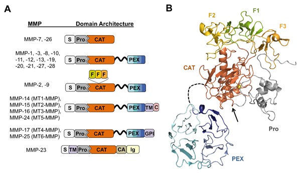
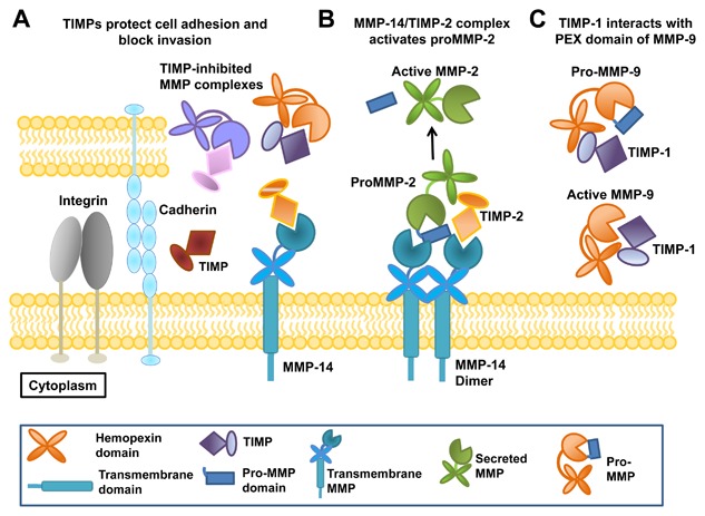
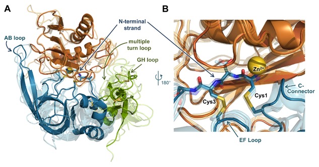
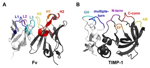
Matrix metalloproteinases (MMPs) are a family of zinc endopeptidases that cleave nearly all components of the extracellular matrix as well as many other soluble and cell-associated proteins. MMPs have been implicated in normal physiological processes, including development, and in the acquisition and progression of the malignant phenotype. Disappointing results from a series of clinical trials testing small molecule, broad spectrum MMP inhibitors as cancer therapeutics led to a re-evaluation of how MMPs function in the tumor microenvironment, and ongoing research continues to reveal that these proteins play complex roles in cancer development and progression. It is now clear that effective targeting of MMPs for therapeutic benefit will require selective inhibition of specific MMPs. Here, we provide an overview of the MMP family and its biological regulators, the tissue inhibitors of metalloproteinases (TIMPs). We then summarize recent research from model systems that elucidate how specific MMPs drive the malignant phenotype of breast cancer cells, including acquisition of cancer stem cell features and induction of the epithelial-mesenchymal transition, and we also outline clinical studies that implicate specific MMPs in breast cancer outcomes. We conclude by discussing ongoing strategies for development of inhibitors with therapeutic potential that are capable of selectively targeting the MMPs most responsible for tumor promotion, with special consideration of the potential of biologics including antibodies and engineered proteins based on the TIMP scaffold. J. Cell. Biochem. 118: 3531-3548, 2017. © 2017 The Authors. Journal of Cellular Biochemistry Published by Wiley Periodicals, Inc.

Keywords: BREAST CANCER; CANCER BIOMARKERS; EPITHELIAL MESENCHYMAL TRANSITION; MATRIX METALLOPROTEINASES; MMP INHIBITORS; TISSUE INHIBITORS OF METALLOPROTEINASES; TUMOR MICROENVIRONMENT; TUMOR PROGRESSION.


Figures

Figure 1
Figure 1
MMP domain structure and protein fold. (A) The domain organization of each human MMP is illustrated schematically; S, signal peptide; Pro, propeptide; CAT, catalytic domain; F, fibronectin type II repeats; PEX, hemopexin domain; TM, transmembrane domain; GPI, glycophosphatidylinositol membrane anchor; C, cytoplasmic domain; CA, cysteine array; Ig, immunoglobulin‐like domain. The flexible, variable length linker between CAT and PEX is shown as a black ribbon. (B) The representative 3D protein fold of proMMP‐2 is illustrated; individual domains are colored as in panel A. The flexible linker between CAT and PEX domains, shown as a black dashed line, varies in length among MMPs. The prodomain (gray) inhibits activity by coordinating the catalytic zinc (yellow sphere) and blocking access to substrates. Activation requires proteolysis within the loop indicated by the black arrow, leading to dissociation of the prodomain. Figure was generated with PyMOL (Schrodinger, LLC) from coordinates of PDB ID: 1GXD [Morgunova et al., 2002].
Figure 2
Figure 2
TIMPs regulate MMP function and activation. (A) Soluble and extracellular matrix‐associated TIMPs form inhibitory interactions with the catalytic domains of extracellular and transmembrane MMPs, protecting cell adhesion and blocking invasion. (B) The dimeric form of transmembrane MMP‐14 forms a receptor complex with TIMP‐2 that is responsible for MMP‐2 activation; proMMP‐2 is bound via its C‐terminal PEX domain, allowing proteolysis of the prodomain by the uninhibited MMP‐14 subunit. (C) TIMP‐1 forms C‐terminal domain interactions with the PEX domain of proMMP‐9 or MMP‐9, protecting proMMP‐9 from activation and quenching activity after activation.
Figure 3
Figure 3
MMP/TIMP interfaces feature a conserved core interaction and diverse peripheral interactions. (A) The structure of the representative complex of MMP‐10CAT (orange) with TIMP‐2 (N‐terminal domain blue, C‐terminal domain green) (PDB ID: 4ILW) [Batra et al., 2013] is shown; the catalytic zinc ion is rendered as a yellow sphere. Four additional MMPCAT/TIMP structures (semi‐transparent) are superposed to highlight regions of structural conservation versus diversity: MMP‐10CAT/TIMP‐1 (PDB ID: 3V96) [Batra et al., 2012a], MMP‐3CAT/TIMP‐1 (PDB ID: 1UEA)[Gomis‐Ruth et al., 1997], MMP‐14CAT/TIMP‐2 (PDB ID: 1BQQ)[Fernandez‐Catalan et al., 1998], and MMP‐13CAT/TIMP‐2 (PDB ID: 2E2D)[Maskos et al., 2007]. Diverse interactions with different MMPs are formed by peripheral TIMP epitopes of the AB, GH, and multiple turn loops. (B) Closer view of the superposed complexes reveals conserved positioning and interactions of the TIMP core epitope, including N‐terminal residues 1–4, the EF loop, and C‐connector loop. The amine terminus of residue Cys‐1 coordinates directly to the catalytic zinc ion. Figure was generated using PyMOL (Schrodinger, LLC).
Figure 4
Figure 4
Similarities between antibody Fv and TIMP binding interfaces. (A) An Fv binds to an antigen through three loops of the heavy chain (dark gray): complementarity‐determining regions (CDRs) H1 (orange), H2 (red), and H3 (yellow), and three loops of the light chain (light gray): CDRs L1 (blue), L2 (purple), and L3 (cyan). Coordinates are from PDB ID: 1YQV [Cohen et al., 2005]. (B) TIMPs, like antibodies, recognize the MMP target using a broad interface comprised of multiple loops spread across two domains, including segments of the N‐terminal domain (dark gray): N‐terminus (purple), AB loop (yellow), C‐connector loop (red), and EF loop (orange), and segments of the C‐terminal domain (light gray): GH loop (cyan) and multiple‐turn loop (blue). Coordinates are from PDB ID: 1UEA [Gomis‐Ruth et al., 1997]. Figure was generated using PyMOL (Schrodinger, LLC).

References

    1. Alvarez OA, Carmichael DF, DeClerck YA. 1990. Inhibition of collagenolytic activity and metastasis of tumor cells by a recombinant human tissue inhibitor of metalloproteinases. J Natl Cancer Inst 82(7):589–595. - PubMed
    1. Annes JP, Munger JS, Rifkin DB. 2003. Making sense of latent TGFbeta activation. J Cell Sci 116(Pt 2):217–224. - PubMed
    1. Amour A, Knight CG, English WR, Webster A, Slocombe PM, Knauper V, Docherty AJ, Becherer JD, Blobel CP, Murphy G. 2002. The enzymatic activity of ADAM8 and ADAM9 is not regulated by TIMPs. FEBS Lett 524(1‐3):154–158. - PubMed
    1. Ardi VC, Kupriyanova TA, Deryugina EI, Quigley JP. 2007. Human neutrophils uniquely release TIMP‐free MMP‐9 to provide a potent catalytic stimulator of angiogenesis. Proc Natl Acad Sci USA 104(51):20262–20267. - PMC - PubMed
    1. Ardi VC, Van den Steen PE, Opdenakker G, Schweighofer B, Deryugina EI, Quigley JP. 2009. Neutrophil MMP‐9 proenzyme, unencumbered by TIMP‐1, undergoes efficient activation in vivo and catalytically induces angiogenesis via a basic fibroblast growth factor (FGF‐2)/FGFR‐2 pathway. J Biol Chem 284(38):25854–25866. - PMC - PubMed

Publication types

MeSH terms

Substances