. 2017 Aug;40(2):293-302.
doi: 10.3892/ijmm.2017.3009. Epub 2017 Jun 2.

Lewis(y) antigen promotes the progression of epithelial ovarian cancer by stimulating MUC1 expression

Affiliations

Lewis(y) antigen promotes the progression of epithelial ovarian cancer by stimulating MUC1 expression

Rui Hou et al. Int J Mol Med. 2017 Aug.

Abstract

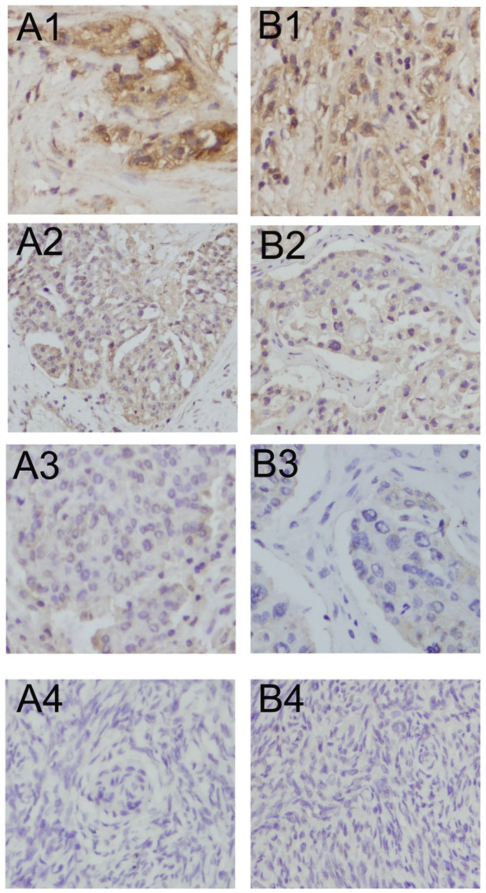
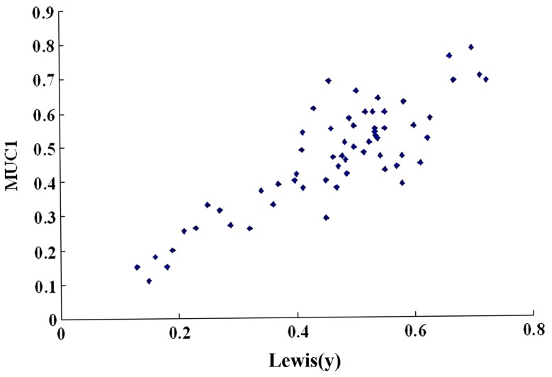
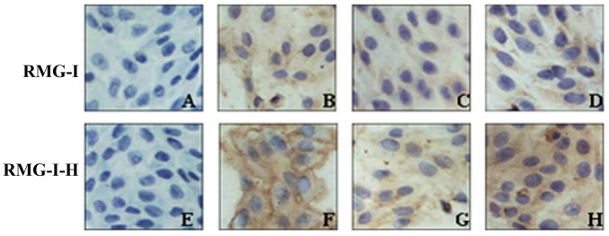
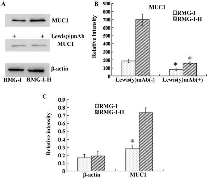
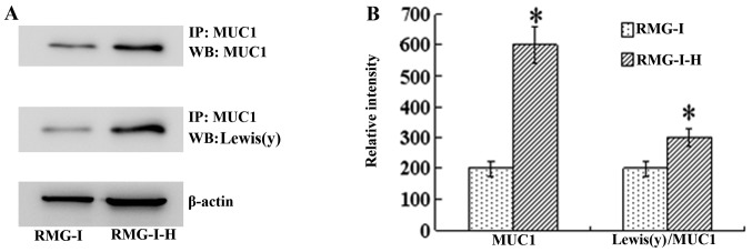
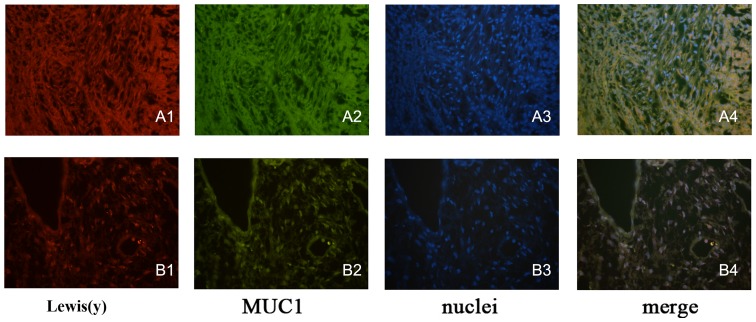
MUC1 is a type I transmembrane glycoprotein and is overexpressed in various epithelial tumor tissues. Some researchers have demonstrated that the glycosylation status of MUC1 can affect MUC1-mediated tumor growth and cell differentiation. In our previous study, we proved that the abilities of cell proliferation, adhesion, invasion and metastasis, and drug resistance were enhanced in ovarian cancer cells stably expressing Lewis(y). Therefore, we hypothesized that Lewis(y) antigen may play a central role in regulating MUC1 expression, and MUC1-mediated cell growth and differentiation may be closely associated with Lewis(y) antigen. This study aimed to examine the correlation between MUC1 expression and Lewis(y) antigen levels in ovarian cancer cell lines and tissue samples. A series of techniques, including RT-qPCR, western blot anlaysis, immunoprecipitation, immunohistochemistry and double-labeling immunofluorescence were applied to detect the expression of Lewis(y) and MUC1. In malignant epithelial ovarian tumors, the positive expression rates of Lewis(y) antigen and MUC1 were 88.33 and 86.67%, respectively, which were markedly higher than those in borderline (60.00 and 53.33%, P<0.05), benign (33.33 and 30%, P<0.01) and normal (0 and 25%, P<0.01) ovarian samples. There was no correlation between the positive expression rates of Lewis(y) or MUC1 and clinicopathological parameters in ovarian cancers (P>0.05). The expression levels of Lewis(y) and MUC1 correlated with the clinical FIGO stage (P<0.05). Both MUC1 and Lewis(y) were highly expressed in ovarian cancer tissues, and their expression levels were positively correlated (P<0.01). In α1,2-fucosyltransferase (α1,2-FT)-transfected cells, the gene and protein expression levels of MUC1 were significantly upregulated compared with the cells that did not overexpress α1,2-FT (P<0.05). The ratio of Lewis(y) immunoprecipitated with MUC1 to total MUC1 increased 1.55-fold in α1,2-FT-overexpressing cells (P<0.05). The overexpression of Lewis(y) resulted in the upregulation of MUC1. On the whole, our data indicate that both MUC1 and Lewis(y) are associated with the occurrence and development of ovarian cancers.


Figures

Figure 1
Figure 1
Immunohistochemical staining of Lewis(y) antigen and MUC1 in various ovarian tissues (×400 magnification). (A1–A4) Lewis(y) antigen expression in ovarian malignant tumor, borderline tumor, benign tumor and normal ovarian tissues, respectively; (B1–B4) MUC1 expression in ovarian malignant tumor, borderline tumor, benign tumor and normal ovarian tissues respectively.
Figure 2
Figure 2
Scatter plot of mean optical density (MOD) value of Lewis(y) antigen and MUC1 in ovarian cancer. The expression intensities of Lewis(y) antigen and MUC1 were linearly correlated (r=0.657, P<0.01).
Figure 3
Figure 3
Immunocytochemical staining of MUC1 in cells before and after α1,2-fucosyltransferase (α1,2-FT) gene transfection (×400 magnification). (A and E) Negative control; (B and F) no treatment; (C and G) pre-treatment with Lewis(y) mAb; (D and H) pre-treatment with irrelevant isotype IgG as control.
Figure 4
Figure 4
Expression of MUC1 proteins in RMG-I and RMG-I-H cells before and after anti-Lewis(y) monoclonal antibody treatment, and the Lewis(y) content of the glycans of MUC1 before and after α1,2-fucosyltransferase (α1,2-FT) gene transfection. (A) Western blots of MUC1 protein in ovarian carcinoma-derived RMG-I and RMG-I-H cells using MUC1 antibody and HRP-labeled secondary antibodies. (B) Densitometric quantification of MUC1 in (A). *P<0.01 compared with RMG-I-H cells without Lewis(y) mAb treatment. Data are presented as the means ± SD (n=3). *P<0.05. (C) RT-qPCR results of mRNA expression of MUC1 of in RMG-I and RMG-I-H cells. *P<0.01 compared with RMG-I-H cells.
Figure 5
Figure 5
Expression of MUC1 and Lewis(y) antigen of the glycans of MUC1 before and after α1,2-fucosyltransferase (α1,2-FT) gene transfection. (A) Western blots of immunoprecipitated MUC1 protein using corresponding antibodies and Lewis(y) antibody and HRP-labeled secondary antibodies. (B) Densitometric quantification of Lewis(y) in (A) and calculation of Lewis(y) expression/MUC1 (set the RMG-I cells as 100%) (n=3). *P<0.01 compared with RMG-I. IP, immunoprecipitation by the antibody of MUC1; WB, western blot with antibodies to MUC1 or Lewis(y). Data are presented as the means ± SD (n=3). *P<0.05.
Figure 6
Figure 6
Lewis(y) and MUC1 co-localize in ovarian cancer tissues (A) and ovarian carcinoma cell RMG-I-H (B) (×400 magnification). Using double-labeling immunofluorescence method, Lewis(y) (A1 and B1), MUC1 (A2 and B2), nuclei stained with DAPI (A3 and B3), merged image [(A4 and B4) co-localization (yellow) of Lewis(y) and MUC1].

References

    1. Goupille C, Hallouin F, Meflah K, Le Pendu J. Increase of rat colon carcinoma cells tumorigenicity by alpha(1-2)fucosyltransferase gene transfection. Glycobiology. 1997;7:221–229. doi: 10.1093/glycob/7.2.221. - DOI - PubMed
    1. Hellström I, Garrigues HJ, Garrigues U, Hellström KE. Highly tumor-reactive, internalizing, mouse monoclonal antibodies to Le(y)-related cell surface antigens. Cancer Res. 1990;50:2183–2190. - PubMed
    1. Madjd Z, Parsons T, Watson NF, Spendlove I, Ellis I, Durrant LG. High expression of Lewis y/b antigens is associated with decreased survival in lymph node negative breast carcinomas. Breast Cancer Res. 2005;7:R780–R787. doi: 10.1186/bcr1305. - DOI - PMC - PubMed
    1. Yang Z, Wu JH, Kuo HW, Kannagi R, Wu AM. Expression of sialyl Lex, sialyl Lea, Lex and Ley glycotopes in secreted human ovarian cyst glycoproteins. Biochimie. 2009;91:423–433. doi: 10.1016/j.biochi.2008.11.002. - DOI - PubMed
    1. Rodríguez-Burford C, Barnes MN, Berry W, Partridge EE, Grizzle WE. Immunohistochemical expression of molecular markers in an avian model: A potential model for preclinical evaluation of agents for ovarian cancer chemoprevention. Gynecol Oncol. 2001;81:373–379. doi: 10.1006/gyno.2001.6191. - DOI - PubMed

MeSH terms