. 2017 Jun 6;12(6):e0179133.
doi: 10.1371/journal.pone.0179133. eCollection 2017.

3D printing and milling a real-time PCR device for infectious disease diagnostics

Affiliations

3D printing and milling a real-time PCR device for infectious disease diagnostics

Geoffrey Mulberry et al. PLoS One. .

Abstract

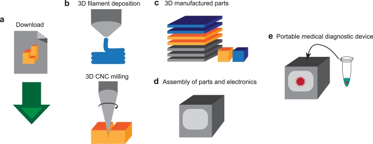
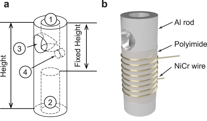
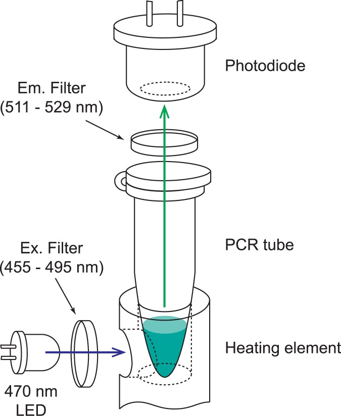
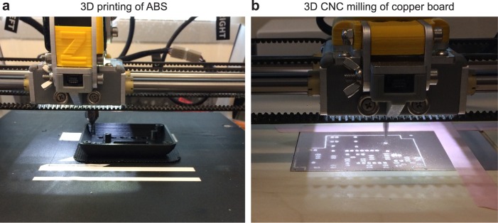
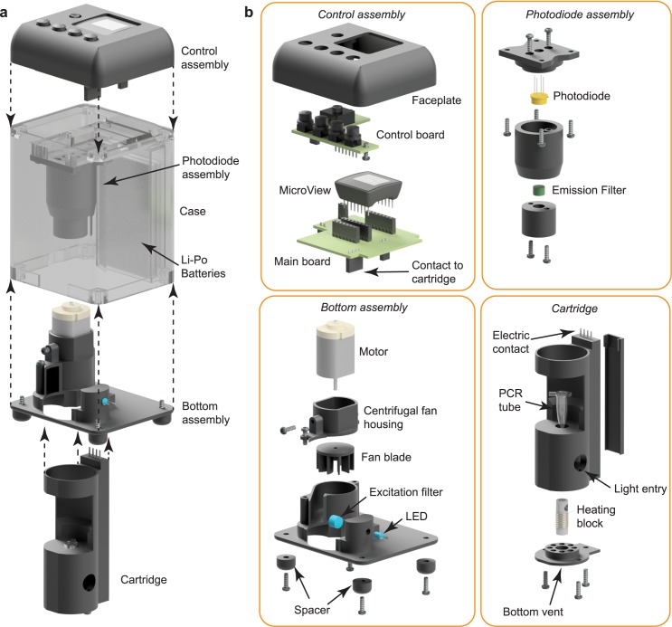
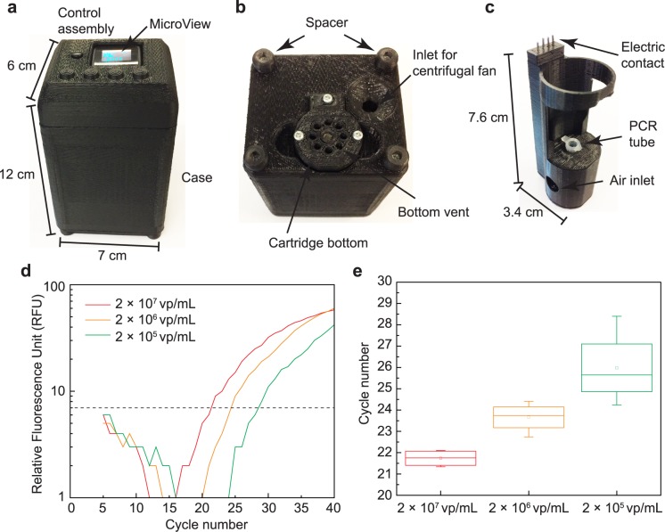
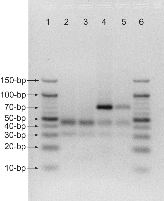
Diagnosing infectious diseases using quantitative polymerase chain reaction (qPCR) offers a conclusive result in determining the infection, the strain or type of pathogen, and the level of infection. However, due to the high-cost instrumentation involved and the complexity in maintenance, it is rarely used in the field to make a quick turnaround diagnosis. In order to provide a higher level of accessibility than current qPCR devices, a set of 3D manufacturing methods is explored as a possible option to fabricate a low-cost and portable qPCR device. The key advantage of this approach is the ability to upload the digital format of the design files on the internet for wide distribution so that people at any location can simply download and feed into their 3D printers for quick manufacturing. The material and design are carefully selected to minimize the number of custom parts that depend on advanced manufacturing processes which lower accessibility. The presented 3D manufactured qPCR device is tested with 20-μL samples that contain various concentrations of lentivirus, the same type as HIV. A reverse-transcription step is a part of the device's operation, which takes place prior to the qPCR step to reverse transcribe the target RNA from the lentivirus into complementary DNA (cDNA). This is immediately followed by qPCR which quantifies the target sequence molecules in the sample during the PCR amplification process. The entire process of thermal control and time-coordinated fluorescence reading is automated by closed-loop feedback and a microcontroller. The resulting device is portable and battery-operated, with a size of 12 × 7 × 6 cm3 and mass of only 214 g. By uploading and sharing the design files online, the presented low-cost qPCR device may provide easier access to a robust diagnosis protocol for various infectious diseases, such as HIV and malaria.


Conflict of interest statement

Competing Interests: The authors have declared that no competing interests exist.

Figures

Fig 1
Fig 1. From digital design file to portable diagnostics device.
(a) A digital format of design file is uploaded to the internet, and downloaded by users. (b) A user uploads the digital files into 3D printer and 3D CNC milling machine for manufacturing. The 3D printer extrudes and deposits filament and the 3D CNC mill cuts and drills through material to engrave patterns and shapes. (c) This results in 3D manufactured parts for assembly. (d) The parts and off-the-shelf electronics components are assembled. (e) This assembled device can function as a portable medical diagnostic device for detecting and quantifying the pathogen in a sample through qPCR.
Fig 2
Fig 2. The aluminum heating element for qPCR device.
(a) The aluminum rod has four holes for ① PCR tube insertion, ② convection cooling, ③ the excitation light path, and ④ thermistor insertion. (b) The heating element consists of an aluminum rod, a layer of polyimide film, and a coil of NiCr wire. The dimension and location of the holes are: ① ⌀ is 3.72 mm and depth is 5.46 mm, ② ⌀ is 3.72 mm and depth is 8.59 mm, ③ ⌀ is 3.56 mm and location from top is 3.70 mm, and ④ ⌀ is 1.59 mm and depth is 1.00 mm and location from top is 3.70 mm.
Fig 3
Fig 3. Optical design of real-time fluorescence measurement for qPCR.
The blue LED (470 nm) emits excitation wavelength that is filtered through an excitation filter which is a band-pass filter of 455 to 495 nm. The excitation light travels through the hole in the aluminum rod and is absorbed to the sample in the PCR tube. The emitted light is collected by an avalanche photodiode through an emission filter, with a band-pass at 511 to 529 nm.
Fig 4
Fig 4. Photographs of 3D printing and 3D milling of all the parts using a multitool 3D printer.
(a) The faceplate is being printed with black ABS material on a heated bed. (b) The main board is being engraved using 3D CNC mill.
Fig 5
Fig 5. 3D assembly of the system with exploded views.
(a) The complete device showing locations of Control assembly, Photodiode assembly, Bottom assembly, Case, and Li-Po batteries. (b) Detail of assemblies. The Control assembly contains a faceplate, a control board, a main board, and a MicroView microcontroller. This assembly provides a user interface and houses all electronic controls. The Photodiode assembly contains the photodiode and emission filter. This assembly is used to determine the target DNA concentration present in the sample. The bottom assembly contains a motor, a fan blade, spacers, and a centrifugal fan housing, forming the cooling fan system. It also contains an LED and excitation filter, establishing the light source for illuminating the sample. The cartridge is a removable assembly allowing for easy insertion of the PCR tube containing the sample. It contains the heating block, electrical contacts for connection to the main board, and a vented bottom plate allowing for the escape of warm air.
Fig 6
Fig 6. The thermal simulation of the heating element’s heating and cooling cycle time.
(a) The heating time from 60°C to 95°C in various heights of heating element. The shortest (9.5 seconds) to heat is 18.5 mm in height. (b) The cooling time from 95°C to 60°C in a fan-operating condition. The shortest (23.9 seconds) to cool is 36.9 mm in height. Increasing the height of the aluminum rod increases the surface area, resulting in improved convection and the length of the cooling period is reduced. The subset graph is an expended view at 23 to 27 seconds. (c) The summary of heating and cooling time for various heights of heating element. The height with the shortest total cycling time is 18.5 mm (36.1 seconds). The heating period increases at a faster rate to the increase in height than the cooling period decreases.
Fig 7
Fig 7. The temperature plot of the sample and the heating element for calibration.
The plot is used to determine proper thermocycling control thresholds. The gray curve shows the temperature of the aluminum heating block; the red curve shows the temperature inside of the 20ul sample inside of the PCR tube.
Fig 8
Fig 8. The photograph of 3D manufactured qPCR device and the resulted RT-qPCR from 3D manufactured qPCR device from three separate qPCR using 3 dilutions of target virus, 2 × 10⁷ vp/mL, 2 × 10⁶ vp/mL, and 2 × 10⁵ vp/mL.
(a) The dimension of the qPCR device is 12 × 7 × 6 cm3. The MicroView shows the status of amplification cycle and fluorescence reading. (b) The bottom view of the qPCR device showing cartridge bottom and an air inlet for centrifugal fan. (c) The size of the cartridge that holds PCR tube during qPCR is 7.6 × 3.4 × 2.9 cm3. (d) Measured fluorescence readings show the shift in the intensity measurements corresponding to the differing concentrations of target virus. The threshold for determining Cq is also shown is a dotted line. (e) Measured Cq for three concentrations of target DNA. The mean and standard deviation of Cq are 21.74 ± 0.39, 23.66 ± 0.70, and 25.98 ± 1.75, for 2 × 10⁷ vp/mL, 2 × 10⁶ vp/mL, and 2 × 10⁵ vp/mL, respectively.
Fig 9
Fig 9. Comparison of conventional qPCR machine and 3D printed device using gel electrophoresis.
Each lane included: (lane 1 and lane 6) 10-bp DNA ladders, (lane 2) negative control from conventional device, (lane 3) negative control from 3D printed device, (lane 4) 2 × 10⁷ vp/mL sample from conventional device, and (lane 5) 2 × 10⁷ vp/mL sample from 3D printed device.

References

    1. Moody A. Rapid diagnostic tests for malaria parasites. Clin Microbiol Rev. American Society for Microbiology; 2002;15: 66–78. doi: 10.1128/CMR.15.1.66-78.2002 - DOI - PMC - PubMed
    1. WHO. Recommended selection criteria for procurement of malaria rapid diagnostic tests. 2016; 16. Available: http://www.who.int/malaria/publications/atoz/rdt-selection-criteria.pdf?...
    1. Rosenberg NE, Kamanga G, Phiri S, Nsona D, Pettifor A, Rutstein SE, et al. Detection of acute HIV infection: a field evaluation of the determine® HIV-1/2 Ag/Ab combo test. J Infect Dis. Oxford University Press; 2012;205: 528–34. doi: 10.1093/infdis/jir789 - DOI - PMC - PubMed
    1. Branson BM, Handsfield HH, Lampe MA, Janssen RS, Taylor AW, Lyss SB, et al. Revised recommendations for HIV testing of adults, adolescents, and pregnant women in health-care settings. MMWR Recomm reports Morb Mortal Wkly report Recomm reports. 2006;55: 1–17–4. Available: http://www.ncbi.nlm.nih.gov/pubmed/16988643 - PubMed
    1. Bloland PB. Drug resistance in malaria. World Heal Organ. 2001; WHO/CDS/CSR/DRS/2001.4. Available: http://www.who.int/csr/resources/publications/drugresist/malaria.pdf

MeSH terms