. 2018 Feb 15;44(2):432-442.
doi: 10.1093/schbul/sbx077.

Isolated Mitochondria Transfer Improves Neuronal Differentiation of Schizophrenia-Derived Induced Pluripotent Stem Cells and Rescues Deficits in a Rat Model of the Disorder

Affiliations

Isolated Mitochondria Transfer Improves Neuronal Differentiation of Schizophrenia-Derived Induced Pluripotent Stem Cells and Rescues Deficits in a Rat Model of the Disorder

Odile Robicsek et al. Schizophr Bull. .

Abstract

Dysfunction of mitochondria, key players in various essential cell processes, has been repeatedly reported in schizophrenia (SZ). Recently, several studies have reported functional recovery and cellular viability following mitochondrial transplantation, mostly in ischemia experimental models. Here, we aimed to demonstrate beneficial effects of isolated active normal mitochondria (IAN-MIT) transfer in vitro and in vivo, using SZ-derived induced pluripotent stem cells (iPSCs) differentiating into glutamatergic neuron, as well as a rodent model of SZ. First, we show that IAN-MIT enter various cell types without manipulation. Next, we show that IAN-MIT transfer into SZ-derived lymphoblasts induces long-lasting improvement in various mitochondrial functions including cellular oxygen consumption and mitochondrial membrane potential (Δ ψ m). We also demonstrate improved differentiation of SZ-derived iPSCs into neurons, by increased expression of neuronal and glutamatergic markers β3-tubulin, synapsin1, and Tbr1 and by an activation of the glutamate-glutamine cycle. In the animal model, we show that intra-prefrontal cortex injection of IAN-MIT in adolescent rats exposed prenatally to a viral mimic prevents mitochondrial Δ ψ m and attentional deficit at adulthood. Our results provide evidence for a direct link between mitochondrial function and SZ-related deficits both in vitro and in vivo and suggest a therapeutic potential for IAN-MIT transfer in diseases with bioenergetic and neurodevelopmental abnormalities such as SZ.


Figures

Fig. 1.
Fig. 1.
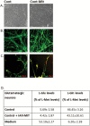
Mitochondrial transfer into healthy and SZ cells. (A) Representative images of time-dependent transfer of JC-1 stained IAN-MIT into SZ-lymphoblasts, (a, b) by time-lapse, (c) confocal microscopy, and (d) JC-1 stained IAN-MIT negative control. (BD) Representative images of JC-1 stained cells (B) 2 weeks after transfer into lymphoblasts, (C) 4 h after transfer into iPSCs, (D) 4 weeks after late IAN-MIT transfer in neurons. (E) PCR products 3 days after IAN-MIT (4 bands) or vehicle (2 bands) with (lanes 1 and 3) and without Alu1 restriction (lanes 2 and 4). (F) Quantification in lymphoblasts of (a) Δ ψ m, (b) mitochondrial distribution, (c) network connectivity. IAN-MIT normalized Δ ψ m and mitochondria distribution impairments, but not network connectivity. (G) Quantification in neurons of (a) Δ ψ m, (b) mitochondrial distribution, and (c) network connectivity. IAN-MIT normalized the impaired Δ ψ m and mitochondria cellular distribution observed in SZ-neurons. JC-1 in cytosol (green) and in active mitochondria (red). Values are mean ± SEM of 2 controls and 2 patients (one iPSCs clone for each) from 2 experiments in duplicates with 5–6 cells in each. Scale bars: A—1 µm (time-lapse) and 3 µm (confocal), B and C—5 µm, D—2 µm. *P < .02, SZ vs Cont; +P < .01, SZ-Mit vs Cont; #P < .03, SZ-Mit vs SZ. (H) IAN-MIT transfer restores abnormal respiration rates and dopamine-induced inhibition in SZ-lymphoblasts 25 days (5 passages) after transfer. aP < .04 and cP < .004 vs control; bP < .03 and dP < .025 vs lymphoblasts + Mit.
Fig. 2.
Fig. 2.
Glutamatergic differentiation following early and late IAN-MIT transfer. (A) Bright-field images of neurons. (B) Immunofluorescence staining and (D) quantification of β3-tubulin and Tbr1, both decreased in SZ neurons. Early and late IAN-MIT transfer increased β3-tubulin, while Tbr1 increased only following late transfer. GLM analysis showed a time-dependent increase in both (β3-tubulin–F = 20.16, P < .001. Tbr1-F = 116.8, P < .001). (C) Immunofluorescence staining and (E) quantification of synapsin1 and PSD-95 and their co-localization, which were decreased in SZ-neurons. Early and Late IAN-MIT transfer had a minor effect on PSD-95 and co-localization, yet profoundly affected synapsin1, which following late transfer reached control levels. GLM showed a time-dependent increase in synapsin1 (Synapsin1-F = 90.15, P < .002). Values are mean ± SEM of 2 controls and 2 patients (one iPSCs clone for each) from 2 experiments in duplicates with 4–6 cells in each. Scale bars: A, B—100 µm, C—25 µm. (F) Late, but not early, IAN-MIT transfer almost normalized Gln release and ameliorated abnormal Glu consumption. Values are mean ± SEM of 2 controls and 2 patients (one iPSCs clone for each) from 2 experiments in triplicates. *P < .01 SZ vs Cont; +P < .008 SZ-Mit vs Cont; #P < .03 SZ-Mit vs SZ.
Fig. 3.
Fig. 3.
IAN-MIT late transfer had no effect on differentiation of control-derived iPSC. (A) Bright field images of neurons. (B) Immunofluorescence staining of β3-tubulin and (C) Tbr1. Scale bar:100 µm. (D) Glu consumption and Gln release in control neurons. Values are mean ± SEM of 2 controls (one iPSCs clone for each) from 2 experiments in duplicates.
Fig. 4.
Fig. 4.
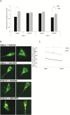
mPFC IAN-MIT transfer prevents disrupted LI and dissipated Δ ψ m in poly-I:C exposed offspring. (A) LI is manifested as shorter log times to complete licking criteria after tone onset of PE compared with NPE groups. Values are mean ± SEM. *P < .0001 PE vs NPE in saline-vehicle exposed offspring and in poly-I:C+IAN-MIT exposed offspring. (B) Representative images of JC-1 stained cortical neurons. JC-1 in cytosol (green), in active mitochondria (red). Scale bar: 5 µm. (C) Quantification of Δ ψ m, which is reduced in poly-I:C rats and increased by IAN-MIT injection in both poly-I:C and saline exposed offspring (n = 4–5 rats/group) *P < .04 IAN-MIT vs Vehicle. #P < .02 Poly-I:C vs Saline.

Similar articles

Cited by

References

    1. Cho B, Choi SY, Cho HM, Kim HJ, Sun W. Physiological and pathological significance of dynamin-related protein 1 (drp1)-dependent mitochondrial fission in the nervous system. Exp Neurobiol. 2013;22:149–157. - PMC - PubMed
    1. Ben-Shachar D, Laifenfeld D. Mitochondria, synaptic plasticity, and schizophrenia. Int Rev Neurobiol. 2004;59:273–296. - PubMed
    1. Sun T, Qiao H, Pan PY, Chen Y, Sheng ZH. Motile axonal mitochondria contribute to the variability of presynaptic strength. Cell Rep. 2013;4:413–419. - PMC - PubMed
    1. Díaz‐Castro B, Pardal R, García‐Flores P, et al. Resistance of glia‐like central and peripheral neural stem cells to genetically induced mitochondrial dysfunction—differential effects on neurogenesis. EMBO Rep. 2015;16:1511–1519. - PMC - PubMed
    1. Rangaraju V, Calloway N, Ryan Timothy A. Activity-driven local ATP synthesis is required for synaptic function. Cell. 2014;156:825–835. - PMC - PubMed

Publication types

MeSH terms