Review
. 2017 Jun 6;7(6):62.
doi: 10.3390/brainsci7060062.

Postnatal Migration of Cerebellar Interneurons

Affiliations
Review

Postnatal Migration of Cerebellar Interneurons

Ludovic Galas et al. Brain Sci. .

Abstract

Due to its continuing development after birth, the cerebellum represents a unique model for studying the postnatal orchestration of interneuron migration. The combination of fluorescent labeling and ex/in vivo imaging revealed a cellular highway network within cerebellar cortical layers (the external granular layer, the molecular layer, the Purkinje cell layer, and the internal granular layer). During the first two postnatal weeks, saltatory movements, transient stop phases, cell-cell interaction/contact, and degradation of the extracellular matrix mark out the route of cerebellar interneurons, notably granule cells and basket/stellate cells, to their final location. In addition, cortical-layer specific regulatory factors such as neuropeptides (pituitary adenylate cyclase-activating polypeptide (PACAP), somatostatin) or proteins (tissue-type plasminogen activator (tPA), insulin growth factor-1 (IGF-1)) have been shown to inhibit or stimulate the migratory process of interneurons. These factors show further complexity because somatostatin, PACAP, or tPA have opposite or no effect on interneuron migration depending on which layer or cell type they act upon. External factors originating from environmental conditions (light stimuli, pollutants), nutrients or drug of abuse (alcohol) also alter normal cell migration, leading to cerebellar disorders.

Keywords: basket cell; cerebellar disorders; cerebellum; drug of abuse; environmental conditions; extracellular matrix; granule cell; interneuron; live-cell imaging; migration; neuropeptides; nutrients; postnatal development; stellate cell.


Conflict of interest statement

The authors declare no conflicts of interest.

Figures

Figure 1
Figure 1
The cortex of the cerebellum: a unique model to study postnatal migration. (A) Complex foliated structure of the postnatal cerebellum with lobules (I–X) as shown in schematic sagittal section. IXa and IXb are sublobules of lobule IX. (B) Interneuron migration: from germinative zones to final location. dWM, deep white matter; pWM, prospective white matter; uRL, upper rhombic lip; EGL, external granular layer; ML, molecular layer; PCL, Purkinje cell layer; IGL, internal granular layer; Gi, Golgi cell; U, unipolar brush cell; G, granule cell; L, Lugaro cell; P, Purkinje cell; B, basket cell; S, stellate cell.
Figure 2
Figure 2
Characteristics of migrating interneurons in the postnatal cerebellum. dWM, deep white matter; pWM, prospective white matter; uRL, upper rhombic lip; EGL, external granular layer; ML, molecular layer; PCL, Purkinje cell layer; IGL, internal granular layer; VZ, ventricular zone; P20, postnatal day 20. * in mouse or rat.
Figure 3
Figure 3
Comparative analysis of imaging technologies used for ex vivo/in vivo interneuron migration studies in the postnatal cerebellum. †, weakly efficient; ††, moderately efficient; †††, highly efficient.
Figure 4
Figure 4
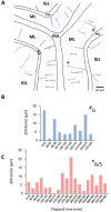
Illustration of the concomitant migration of granule and basket/stellate cells in the postnatal cerebellum through confocal macroscopy. (A) Tracking of centripetal migration (towards the bottom of the IGL) of granule cells (blue) and centrifugal migration (towards the top of ML) of basket/stellate cells (pink). (B) Saltatory movements of a granule cell (marked as # in (A)) in the ML. (C) Saltatory movements of a basket/stellate cell (marked as * in (A)) in the ML. EGL, external granular layer; ML, molecular layer; PCL, Purkinje cell layer; IGL, internal granular layer.
Figure 5
Figure 5
Transient arrest phase, standby phase and stop signals for interneurons during migration in the postnatal cerebellar cortex. pWM, prospective white matter; EGL, external granular layer; ML, molecular layer; PCL, Purkinje cell layer; IGL, internal granular layer; Gi, Golgi cell; U, unipolar brush cell; G, granule cell; L, Lugaro cell; P, Purkinje cell; B, basket cell; S, stellate cell.
Figure 6
Figure 6
Multifactorial control of interneuron migration in the postnatal cerebellar cortex. Cortical-layer-specific effects of molecules that facilitate (in blue), stimulate (in green) or inhibit (in red) cell movements. pWM, prospective white matter; EGL, external granular layer; ML, molecular layer; PCL, Purkinje cell layer; IGL, internal granular layer; Gi, Golgi cell; U, unipolar brush cell; G, granule cell; L, Lugaro cell; P, Purkinje cell; B, basket cell; S, stellate cell; tPA, tissue-type plasminogen activator; PACAP: pituitary adenylate cyclase-activating polypeptide; NT-3, neurotrophin-3; MMP, matrix metalloproteinase; IGF-1, insulin growth factor-1; SDF-1α, stromal cell-derived factor-1α; BDNF, brain-derived nerve factor; PAF, platelet-activating factor.

Similar articles

Cited by

References

    1. Ramón y Cajal S. Cervelet, cerveau moyen, rétine, couche optique, corps strié, écorce cérébrale générale et régionale, grand sympathique. In: Azoulay L., editor. Histologie du Système Nerveux de L’homme et des Vertébrés. 1st ed. Volume 2 Maloine A.; Paris, France: 1911.
    1. Miale I.L., Sidman R.L. An autoradiographic analysis of histogenesis in the mouse cerebellum. Exp. Neurol. 1961;4:277–296. doi: 10.1016/0014-4886(61)90055-3. - DOI - PubMed
    1. Rakic P. Neuron-glia relationship during granule cell migration in developing cerebellar cortex. A Golgi and electonmicroscopic study in Macacus rhesus. J. Comp. Neurol. 1971;141:283–312. doi: 10.1002/cne.901410303. - DOI - PubMed
    1. Edmondson J.C., Hatten M.E. Glial-guided granule neuron migration in vitro: A high-resolution time-lapse video microscopic study. J. Neurosci. 1987;78:1928–1934. - PMC - PubMed
    1. Sillitoe R.V., Joyner A.L. Morphology, molecular codes, and circuitry produce the three-dimensional complexity of the cerebellum. Annu. Rev. Cell. Dev. Biol. 2007;23:549–577. doi: 10.1146/annurev.cellbio.23.090506.123237. - DOI - PubMed

LinkOut - more resources