. 2017 Jun;13(6):2691-2698.
doi: 10.3892/etm.2017.4370. Epub 2017 Apr 20.

Three-dimensional arterial spin labeling imaging and dynamic susceptibility contrast perfusion-weighted imaging value in diagnosing glioma grade prior to surgery

Affiliations

Three-dimensional arterial spin labeling imaging and dynamic susceptibility contrast perfusion-weighted imaging value in diagnosing glioma grade prior to surgery

Hong Ma et al. Exp Ther Med. 2017 Jun.

Abstract

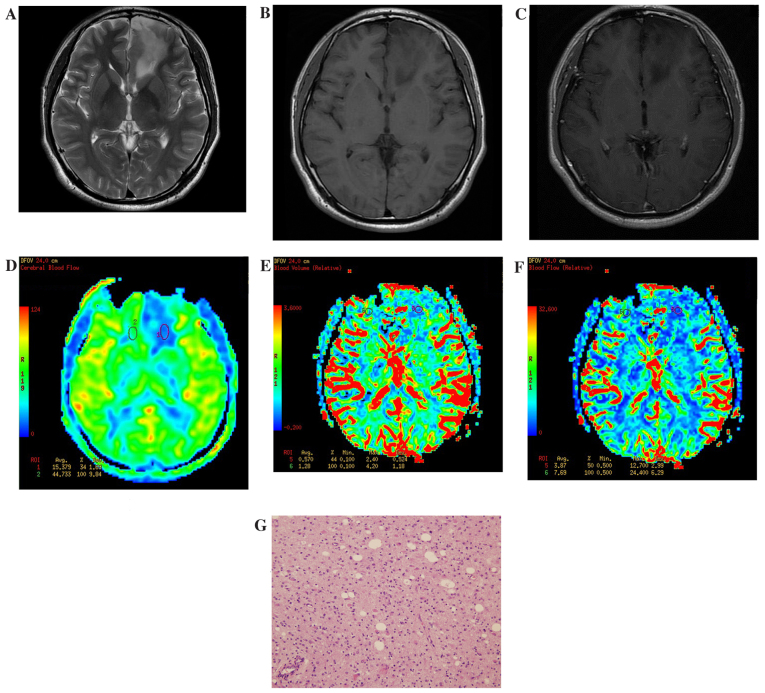
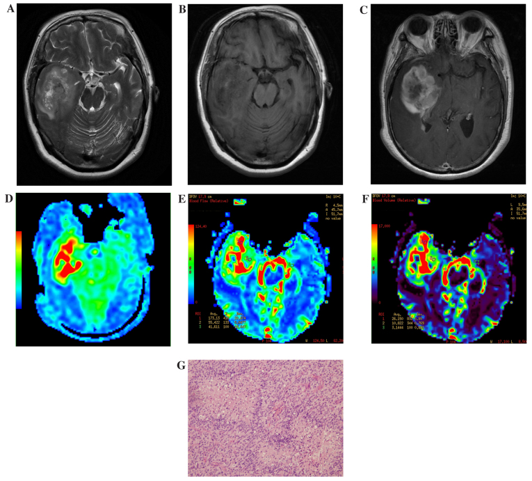
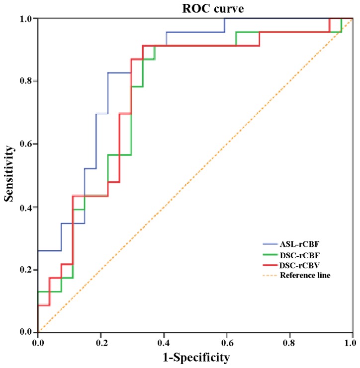
The current study aimed to investigate whole-brain three-dimensional arterial spin labeling imaging (3D ASL) and dynamic susceptibility contrast perfusion-weighted imaging (DSC-PWI), in regards to their diagnostic value of preoperative glioma grade. The parameter values obtained after correction will be correlated with the diagnostic value of 3D ASL and DSC-PWI perfusion. In the current study, 50 patients with gliomas confirmed by pathology were used, including 27 low-grade gliomas (LGGs) and 23 high-grade gliomas (HGGs). Prior to surgery all patients underwent 3 Tesla magnetic resonance imaging (MRI), 3D ASL, DSC-PWI and conventional enhanced MRI scans to obtain original 3D ASL and DSC-PWI images, and the tumor regions with the most obvious parenchyma perfusion and contralateral normal white matter were selected. In these areas, the ASL-relative cerebral blood flow (ASL-rCBF), DSC-relative cerebral blood flow (DSC-rCBF) and DSC-relative cerebral blood volume (DSC-rCBV) parameter values were then obtained after correction for individual differences. The results of the present study show that ASL-CBF, DSC-CBF, DSC-CBV values and ASL-rCBF, DSC-rCBF, DSC-rCBV values increased as the grade of the glioma being imaged increased, and there was a marked difference between the HGGs and the LGGs. ASL-rCBF was significantly positively correlated with DSC-rCBF (r=0.580, P<0.01). In addition, ASL-rCBF was significantly positively correlated with DSC-rCBV (r=0.431, P<0.01). Receiver operating characteristic (ROC) curves were applied to compare the two perfusion parameters of DSC-PWI and 3D ASL in the diagnosis of glioma grade. ASL-rCBF had the highest area value under the ROC curve (0.836). The areas under the ROC curve of DSC-rCBF and DSC-rCBV were analyzed using the Z test, but the difference was not statistically significant. When ASL-rCBF, DSC-rCBF and DSC-rCBV were cutoff at 2.24, 1.85 and 1.68, the sensitivity of HGG diagnosis was 83.2, 91.3 and 91.3%, and the specificity was 77.7, 63.9 and 66.7%, respectively.

Keywords: dynamic susceptibility contrast perfusion-weighted imaging; glioma; magnetic resonance imaging; three-dimensional arterial spin labeling.


Figures

Figure 1.
Figure 1.
Results of imaging of a patient with a low-grade glioma located at the left side of the frontal lobe. (A) Routine T2WI. The focal lesion showed a slightly higher signal. (B) Routine T1WI. The focal lesion showed a slightly low signal. (C) Enhanced T1WI. The focal lesion had no obvious enhancement. (D) ASL-CBF imaging. Low perfusion of the tumor was observed, with a ASL-rCBF value of 0.46. (E) DSC-CBV imaging. Low perfusion of the tumor was observed, with a DSC-rCBV value of 0.44. (F) DSC-CBF imaging. Low perfusion of the tumor was observed, with a DSC-rCBF value of 0.5. (G) Hematoxylin and eosin staining of a low-grade glioma specimen (magnification, ×40). The volume of tumor cells was large with abundant cytoplasm. WI, weighted imaging; ASL, arterial spin labelling; CBF, cerebral blood flow; DSC, dynamic susceptibility contrast; CBV, cerebral blood volume; r, relative.
Figure 2.
Figure 2.
Results of imaging of a patient with a high-grade glioma located at the right side of the temporal lobe. (A) Routine T2WI. The mass showed a slightly higher and higher hybrid signal. The ring pool and saddle pool exhibited pressured narrowing. (B) Routine T1WI. The majority of the mass demonstrated uneven low signal mixed with high signal small patch sample bleeding. (C) Enhanced T1WI. The majority of the mass demonstrated obvious uneven reinforcement and the center area presented low signal without reinforcement. (D) ASL-CBF imaging. Visible and uneven high perfusion was observed, with a ASL-rCBF value of 8.03. (E) DSC-CBV imaging. The mass showed visible and uneven high perfusion, with a DSC-rCBV value of 8.16. (F) DSC-CBF imaging. The mass showed visible and uneven high perfusion, with a DSC-rCBF value of 4.16. (G) Hematoxylin and eosin staining of a high-grade glioma specimen (magnification, ×40). Hyperplasia of the capillaries was observed and vascular endothelial cell enlargement and an increase number of vascular endothelial cells was evident. WI, weighted imaging; ASL, arterial spin labelling; CBF, cerebral blood flow; DSC, dynamic susceptibility contrast; CBV, cerebral blood volume; r, relative.
Figure 3.
Figure 3.
ROC curves of 3D ASL and dynamic susceptibility contrast perfusion-weighted imaging. 3D ASL relative cerebral blood flow had the largest area under the ROC curve, which was 0.836. ROC, receiver operating characteristic. 3D ASL, three-dimensional arterial spin labeling; rCBF, relative cerebral blood flow; DSC, dynamic susceptibility contrast perfusion-weighted imaging; rCBV, relative cerebral blood volume.

Similar articles

Cited by

References

    1. Fuller GN, Scheithauer BW. The 2007 Revised World Health Organization (WHO) Classification of Tumours of the Central Nervous System: Newly codified entities. Brain pathology. 2007;17:304–307. doi: 10.1111/j.1750-3639.2007.00084.x. - DOI - PMC - PubMed
    1. Louis DN, Ohgaki H, Wiestler OD, Cavenee WK, Burger PC, Jouvet A, Scheithauer BW, Kleihues P. The 2007 WHO classification of tumours of the central nervous system. Acta neuropathologica. 2007;114:97–109. doi: 10.1007/s00401-007-0278-6. - DOI - PMC - PubMed
    1. Brat DJ, Parisi JE, Kleinschmidt-DeMasters BK, Yachnis AT, Montine TJ, Boyer PJ, Powell SZ, Prayson RA, McLendon RE. Neuropathology Committee, College of American Pathologists: Surgical neuropathology update: A review of changes introduced by the WHO classification of tumours of the central nervous system, 4th edition. Arch Pathol Lab Med. 2008;132:993–1007. - PubMed
    1. Sulman EP, Guerrero M, Aldape K. Beyond grade: Molecular pathology of malignant gliomas. Semin Radiat Oncol. 2009;19:142–149. doi: 10.1016/j.semradonc.2009.02.001. - DOI - PubMed
    1. Gupta RK, Awasthi R, Rathore RK, Verma A, Sahoo P, Paliwal VK, Prasad KN, Pandey CM, Narayana PA. Understanding epileptogenesis in calcified neurocysticercosis with perfusion MRI. Neurology. 2012;78:618–625. doi: 10.1212/WNL.0b013e318248deae. - DOI - PubMed