

A case report of the orbit, ocular association and the lung in granulomatosis with polyangiitis: A diagnostic challenge
- PMID: 28587410
- PMCID: PMC5450685
- DOI: 10.3892/etm.2017.4408
A case report of the orbit, ocular association and the lung in granulomatosis with polyangiitis: A diagnostic challenge
Abstract
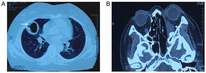
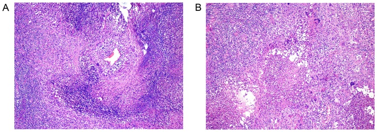
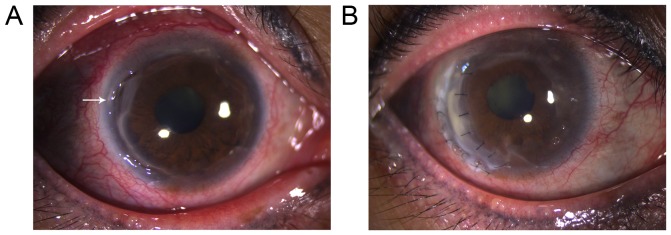
Granulomatosis with polyangiitis (GPA) is a systemic form of vasculitis that involves small to medium sized vessels and is associated with high morbidity and mortality. GPA presents a continuous and difficult clinical diagnostic concern, due to the rarity of the disease and the diversity of the manifestations. This case report discusses the unusual symptoms presented by a particular patient, discusses these manifestations and explains how the final diagnosis was identified as GPA. A 40-year old Chinese woman was initially referred to the present institution for a progressive worsening pain, redness and gradual decrease in visual acuity in the eyes over the past 7-year period. Previous therapeutic interventions included noncompliant topical and intravenous dexamethasone for 6 years. A pre-operative examination conducted in a differing hospital to search for the presence of an orbital mass, resulted in the identification of an asymptomatic space-occupying lesion in the right middle lung, which was surgically removed in March 2015. A total of four weeks later, surgery was then applied to remove a left orbital mass, in the same hospital. A total of three months later, the patient was diagnosed with peripheral ulcerative keratitis associated with GPA, at the present institution. The corneal lesions were then treated bilaterally with cryotherapy and oral prednisone and cyclophosphamide were administered. Following surgery, the condition of the eyes appeared to be stable. A total of seven months later, the redness and pain of the right eye recurred, followed by a deep lamellar keratoplasty for the treatment of necrotizing scleritis. The condition of the two eyes was subsequently observed to be stable during the nine month follow-up. The present case study reviews various points to consider in a rare, complicated and potentially blinding case of GPA. GPA must therefore be considered in the differential diagnosis of further inflammatory conditions and tumors. Early diagnosis and an appropriate interdisciplinary approach to management, are required to decrease recurrence and morbidity in patients with GPA-mediated inflammatory ocular disease.
Keywords: granulomatosis with polyangiitis; lung lesions; orbital mass; peripheral ulcerative keratitis.
Figures
Similar articles
-
Surgical treatment of peripheral ulcerative keratitis and necrotizing scleritis in granulomatosis with polyangiitis.Saudi Med J. 2016 Feb;37(2):205-7. doi: 10.15537/smj.2016.2.13390. Saudi Med J. 2016. PMID: 26837405 Free PMC article.
-
Ocular manifestations of granulomatosis with polyangiitis: report from a tertiary eye care center.Int Ophthalmol. 2024 Feb 7;44(1):16. doi: 10.1007/s10792-024-02989-y. Int Ophthalmol. 2024. PMID: 38321188
-
Case report peripheral ulcerative keratitis with nodular scleritis, presents as the primary manifestation of granulomatosis with polyangiitis.Ann Med Surg (Lond). 2023 May 23;85(7):3658-3662. doi: 10.1097/MS9.0000000000000871. eCollection 2023 Jul. Ann Med Surg (Lond). 2023. PMID: 37427242 Free PMC article.
-
Severe ophthalmic manifestation in pituitary-involved granulomatosis with polyangiitis: a case report and literature review.BMC Ophthalmol. 2018 Nov 16;18(1):299. doi: 10.1186/s12886-018-0966-0. BMC Ophthalmol. 2018. PMID: 30445952 Free PMC article. Review.
-
[Ophthalmologic manifestation of granulomatosis with polyangiitis (Wegener's granulomatosis)].Ter Arkh. 2015;87(12):111-116. doi: 10.17116/terarkh20158712111-116. Ter Arkh. 2015. PMID: 27022660 Review. Russian.
Cited by
-
A Case of an Elderly Woman Who Developed Corneal Perforation in the Clinical Course of Myeloperoxidase Positive Antineutrophil Cytoplasmic Antibody-Associated Vasculitis.Case Rep Rheumatol. 2023 Aug 26;2023:4246075. doi: 10.1155/2023/4246075. eCollection 2023. Case Rep Rheumatol. 2023. PMID: 37662600 Free PMC article.
References
-
- Mahr AD, Neogi T, Merkel PA. Epidemiology of Wegener's granulomatosis: Lessons from descriptive studies and analyses of genetic and environmental risk determinants. Clin Exp Rheumatol. 2006;24(2 Suppl 41):S82–S91. - PubMed
-
- Bohm M, Fernandez MI Gonzalez, Ozen S, Pistorio A, Dolezalova P, Brogan P, Barbano G, Sengler C, Klein-Gitelman M, Quartier P, et al. Clinical features of childhood granulomatosis with polyangiitis (wegener's granulomatosis) Pediatr Rheumatol Online J. 2014;12:18. doi: 10.1186/1546-0096-12-18. - DOI - PMC - PubMed
-
- Chua J, Lim L. Systemic Wegener's granulomatosis with severe orbito-ocular involvement. Singapore Med J. 2008;49:e259–e262. - PubMed
-
- Langford CA, Sneller MC. Update on the diagnosis and treatment of Wegener's granulomatosis. Adv Intern Med. 2001;46:177–206. - PubMed
LinkOut - more resources
Full Text Sources
Other Literature Sources
Miscellaneous