

ERK1/2 pathway regulates coxsackie and adenovirus receptor expression in mouse cardiac stem cells
- PMID: 28587412
- PMCID: PMC5450675
- DOI: 10.3892/etm.2017.4414
ERK1/2 pathway regulates coxsackie and adenovirus receptor expression in mouse cardiac stem cells
Abstract
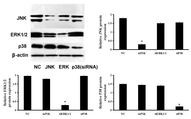
Cardiac stem cells (CSCs) are the most promising and effective candidates for the therapy of cardiac regenerative diseases; however, they have marked limitations. For instance, the implantation of CSCs is hampered by factors such as their sustainability and long-term durability. Gene modification appears to be the most effective method of optimizing CSCs and gene therapy trials have demonstrated that efficient gene transfer is key to achieving therapeutic efficacy. However, the transduction ability of adenovirus (Ad) is limited. Previous studies have reported that low expression of coxsackie and adenovirus receptor (CAR) in target cells decreases the transduction efficiency. A promising method for improving Ad-mediated gene transfer is to increase CAR expression in target cells. The present study investigated the effect of the Raf-mitogen-associated protein kinase (MAPK) kinase (MEK)-extracellular signal-associated protein kinase (ERK) signaling pathway on the expression of CAR on CSCs, as this pathway decreases cell-cell adhesion via cell surface molecules. The results demonstrated that interference with the Raf-MEK-ERK signaling pathway by knockdown of ERK1/2 upregulated the expression of CAR. The entry of the Ad into the cells was increased following inhibition of ERK1/2. Moreover, following knockdown of CAR, the entry of Ad into cells was decreased. However, knockdown of c-Jun N-terminal kinase and p38 as other components of the MAPK pathway did not affect CAR expression. Therefore, CAR expression in CSCs may be mediated via the Raf-MEK-ERK signaling pathway. Upregulation of CAR by knockdown of ERK1/2 may significantly improve Ad-mediated genetic modification of CSCs in the treatment of cardiovascular diseases.
Keywords: cardiac stem cells; coxsackie and adenovirus receptor; extracellular signal-regulated kinase 1/2; gene therapy.
Figures


Similar articles
-
Inhibition of the Raf/MEK/ERK pathway up-regulates expression of the coxsackievirus and adenovirus receptor in cancer cells.Cancer Res. 2003 May 1;63(9):2088-95. Cancer Res. 2003. PMID: 12727824
-
Modification of the genetic program of human alveolar macrophages by adenovirus vectors in vitro is feasible but inefficient, limited in part by the low level of expression of the coxsackie/adenovirus receptor.Am J Respir Cell Mol Biol. 1999 Mar;20(3):361-70. doi: 10.1165/ajrcmb.20.3.3398. Am J Respir Cell Mol Biol. 1999. PMID: 10030833
-
ERK1/2 and MEK1/2 induced by Kaposi's sarcoma-associated herpesvirus (human herpesvirus 8) early during infection of target cells are essential for expression of viral genes and for establishment of infection.J Virol. 2005 Aug;79(16):10308-29. doi: 10.1128/JVI.79.16.10308-10329.2005. J Virol. 2005. PMID: 16051824 Free PMC article.
-
Targeting the Raf-MEK-ERK mitogen-activated protein kinase cascade for the treatment of cancer.Oncogene. 2007 May 14;26(22):3291-310. doi: 10.1038/sj.onc.1210422. Oncogene. 2007. PMID: 17496923 Review.
-
Targeting ERK1/2 protein-serine/threonine kinases in human cancers.Pharmacol Res. 2019 Apr;142:151-168. doi: 10.1016/j.phrs.2019.01.039. Epub 2019 Feb 20. Pharmacol Res. 2019. PMID: 30794926 Review.
Cited by
-
Oncolytic virotherapy against lung cancer: key receptors and signaling pathways of viral entry.Front Immunol. 2024 Oct 4;15:1473288. doi: 10.3389/fimmu.2024.1473288. eCollection 2024. Front Immunol. 2024. PMID: 39430750 Free PMC article. Review.
References
LinkOut - more resources
Full Text Sources
Other Literature Sources
Research Materials
Miscellaneous