

Lymph node tuberculosis mimicking malignancy on 18F-FDG PET/CT in two patients: A case report
- PMID: 28587415
- PMCID: PMC5450601
- DOI: 10.3892/etm.2017.4421
Lymph node tuberculosis mimicking malignancy on 18F-FDG PET/CT in two patients: A case report
Abstract
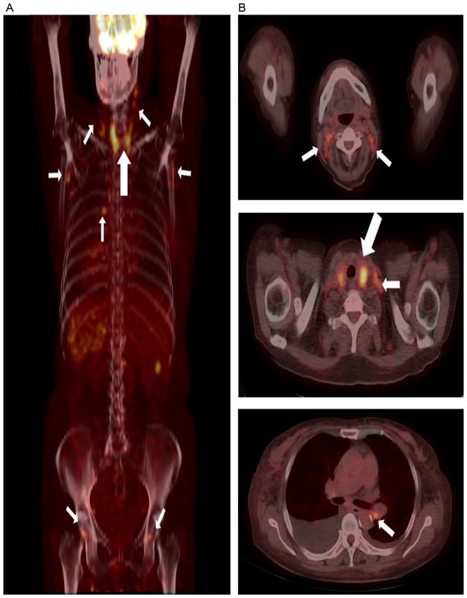
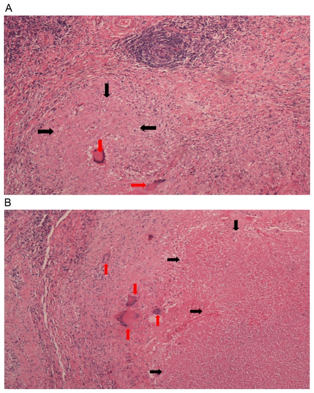
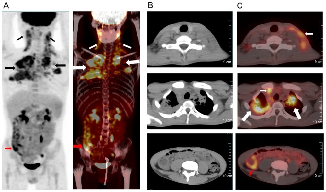
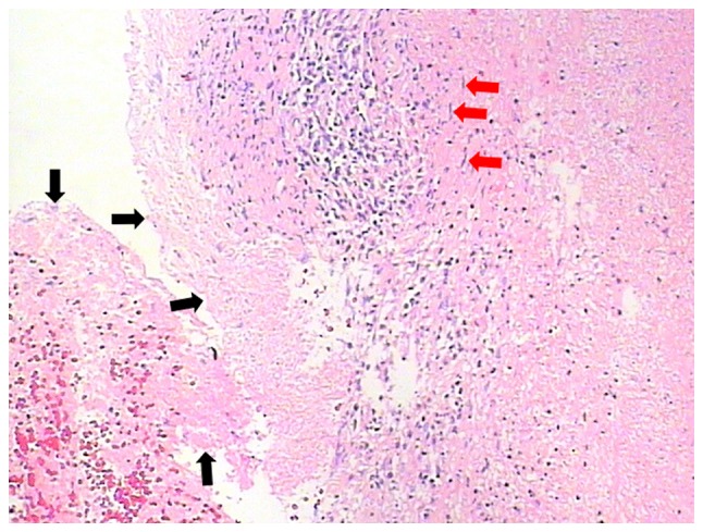
18F-fluorodeoxyglucose positron emission/computed tomography (18F-FDG PET/CT) imaging, an established procedure for evaluation of malignancy, reports an increased 18F-FDG uptake in acute or chronic inflammatory condition. Lymph node tuberculosis (LNTB) is the most common form of extrapulmonary tuberculosis. However, the absence of clinical symptoms and bacteriological basis makes it difficult to diagnose. In the current case report, two patients with LNTB mimicking malignant lymphoma are presented by 18F-FDG PET/CT. The objective of the present report is to emphasize that LNTB should be considered as a noteworthy differential diagnosis in patients with enlarged lymph nodes, particularly in tuberculosis-endemic countries, and that lymph node biopsy serves a vital role in diagnosing LNTB.
Keywords: false positive; lymph node tuberculosis; malignancy; positron emission/computed tomography.
Figures
Similar articles
-
Clinical usefulness of 18F-FDG PET/CT for initial staging and assessment of treatment efficacy in patients with lymph node tuberculosis.Nucl Med Biol. 2017 Jul;50:17-24. doi: 10.1016/j.nucmedbio.2017.04.003. Epub 2017 Apr 8. Nucl Med Biol. 2017. PMID: 28426991
-
[Characteristics of extrapulmonary tuberculosis in (18)F-2-fluoro-2-deoxy-D-glucose positron emission tomography-computed tomography: experience from 39 cases].Zhonghua Jie He He Hu Xi Za Zhi. 2012 Mar;35(3):184-8. Zhonghua Jie He He Hu Xi Za Zhi. 2012. PMID: 22781150 Chinese.
-
(18)F-FDG and (18)F-FLT PET/CT imaging in the characterization of mediastinal lymph nodes.Ann Nucl Med. 2016 Apr;30(3):207-16. doi: 10.1007/s12149-015-1047-6. Epub 2015 Dec 11. Ann Nucl Med. 2016. PMID: 26661845 Clinical Trial.
-
Advances in imaging of tuberculosis: the role of ¹⁸F-FDG PET and PET/CT.Curr Opin Pulm Med. 2014 May;20(3):287-93. doi: 10.1097/MCP.0000000000000043. Curr Opin Pulm Med. 2014. PMID: 24614238 Review.
-
Dual-time point 18F-FDG-PET and PET/CT for Differentiating Benign From Malignant Musculoskeletal Lesions: Opportunities and Limitations.Semin Nucl Med. 2017 Jul;47(4):373-391. doi: 10.1053/j.semnuclmed.2017.02.009. Epub 2017 Apr 19. Semin Nucl Med. 2017. PMID: 28583277 Review.
Cited by
-
18F-FAPI PET/CT performs better in evaluating mediastinal and hilar lymph nodes in patients with lung cancer: comparison with 18F-FDG PET/CT.Eur J Med Res. 2024 Jan 3;29(1):9. doi: 10.1186/s40001-023-01494-9. Eur J Med Res. 2024. PMID: 38173034 Free PMC article.
References
-
- Metser U, Even-Sapir E. Increased (18)F-fluorodeoxyglucose uptake in benign, nonphysiologic lesions found on whole-body positron emission tomography/computed tomography (PET/CT): Accumulated data from four years of experience with PET/CT. Semin Nucl Med. 2007;37:206–222. doi: 10.1053/j.semnuclmed.2007.01.001. - DOI - PubMed
-
- Burdick MJ, Stephans KL, Reddy CA, Djemil T, Srinivas SM, Videtic GM. Maximum standardized uptake value from staging FDG-PET/CT does not predict treatment outcome for early-stage non-small-cell lung cancer treated with stereotactic body radiotherapy. Int J Radiat Oncol Biol Phys. 2010;78:1033–1039. doi: 10.1016/j.ijrobp.2009.09.081. - DOI - PubMed
LinkOut - more resources
Full Text Sources
Other Literature Sources