

Diagnostic accuracy of diffusion-weighted whole-body imaging with background body signal suppression/T2-weighted image fusion for the detection of abdominal solid cancer
- PMID: 28587434
- PMCID: PMC5450632
- DOI: 10.3892/etm.2017.4432
Diagnostic accuracy of diffusion-weighted whole-body imaging with background body signal suppression/T2-weighted image fusion for the detection of abdominal solid cancer
Abstract
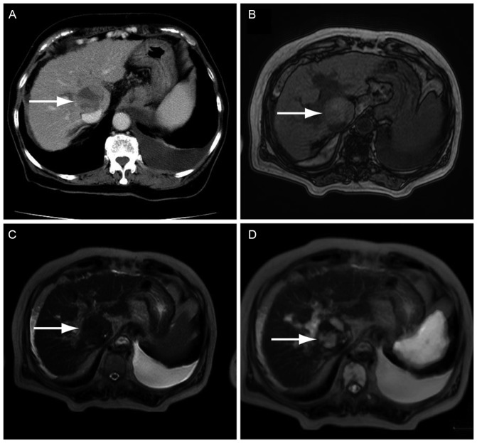
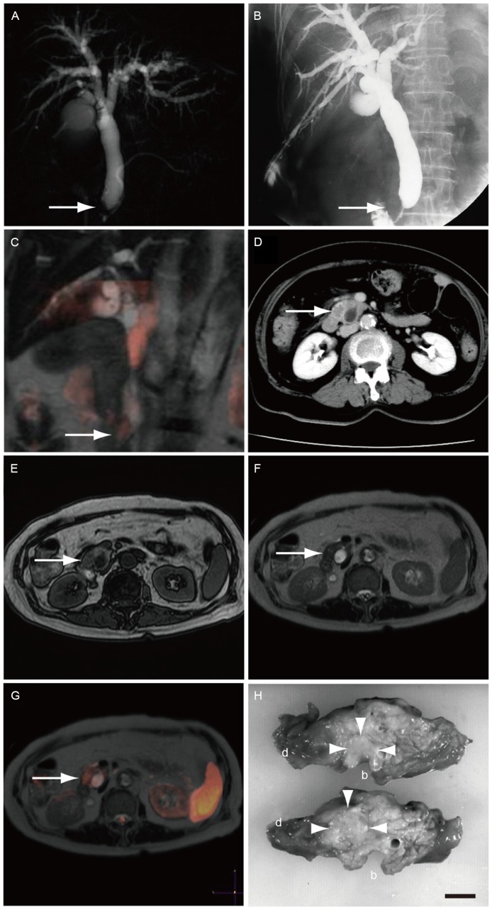
Diffusion-weighted whole-body imaging with background body signal suppression (DWIBS) images show significant contrast for cancer tissues against non-cancerous tissues. Fusion of a DWIBS and a T2-weighted image (DWIBS/T2) can be used to obtain functional, as well as anatomic, information. In the present study, the performance of DWIBS/T2 in the diagnosis of abdominal solid cancer was evaluated. The records of 14 patients were retrospectively analyzed [5 patients with hepatocellular carcinoma (HCC), 4 with metastatic liver cancer, 3 with pancreatic cancer, 1 with renal cellular carcinoma and 1 with malignant lymphoma of the para-aortic lymph node]. T1WI and T2WI scans did not detect pancreatic cancer in certain cases, whereas DWIs and DWIBS/T2 clearly demonstrated pancreatic cancer in all cases. In addition, metastatic liver cancer and HCC were successfully detected with abdominal US and CECT; however, US did not detect pancreatic cancer in 1 case, while CECT and DWIBS/T2 detected pancreatic cancer in all cases. In conclusion, the diagnostic performance of DWIBS/T2 was the same as that of abdominal US and CECT in detecting primary and metastatic liver cancer. DWIBS/T2 enabled the diagnosis of pancreatic cancer in cases where it was not detected with US, T1WI or T2WI.
Keywords: abdominal ultrasonography; computed tomography; hepatocellular carcinoma; pancreatic cancer.
Figures
Similar articles
-
Comparison of DWIBS/T2 image fusion and PET/CT for the diagnosis of cancer in the abdominal cavity.Exp Ther Med. 2017 Oct;14(4):3754-3760. doi: 10.3892/etm.2017.4987. Epub 2017 Aug 22. Exp Ther Med. 2017. PMID: 29042975 Free PMC article.
-
Diffusion-weighted whole body imaging with background body signal suppression/T2 image fusion is negative for patients with intraductal papillary mucinous neoplasm.Hepatogastroenterology. 2015 Mar-Apr;62(138):463-5. Hepatogastroenterology. 2015. PMID: 25916083
-
Diffusion-weighted whole-body imaging with background body signal suppression/T2-weighted image fusion of gastrointestinal cancers.Mol Clin Oncol. 2016 Jul;5(1):44-48. doi: 10.3892/mco.2016.897. Epub 2016 May 11. Mol Clin Oncol. 2016. PMID: 27330763 Free PMC article.
-
[High field magnetic resonance background suppression diffusion imaging in the diagnosis of liver foci of space occupying lesion].Zhong Nan Da Xue Xue Bao Yi Xue Ban. 2013 Mar;38(3):237-44. doi: 10.3969/j.issn.1672-7347.2013.03.004. Zhong Nan Da Xue Xue Bao Yi Xue Ban. 2013. PMID: 23545819 Chinese.
-
Assessing extracranial tumors using diffusion-weighted whole-body MRI.Z Med Phys. 2011 May;21(2):79-90. doi: 10.1016/j.zemedi.2010.06.004. Epub 2010 Oct 2. Z Med Phys. 2011. PMID: 20888200 Review.
Cited by
-
The Influence of Bowel Preparation on ADC Measurements: Comparison between Conventional DWI and DWIBS Sequences.Medicina (Kaunas). 2019 Jul 21;55(7):394. doi: 10.3390/medicina55070394. Medicina (Kaunas). 2019. PMID: 31330916 Free PMC article.
-
The diagnostic performance of contrast-enhanced CT versus extracellular contrast agent-enhanced MRI in detecting hepatocellular carcinoma: direct comparison and a meta-analysis.Abdom Radiol (NY). 2022 Jun;47(6):2057-2070. doi: 10.1007/s00261-022-03484-7. Epub 2022 Mar 21. Abdom Radiol (NY). 2022. PMID: 35312822 Review.
References
-
- Takahara T, Imai Y, Yamashita T, Yasuda S, Nasu S, Van Cauteren M. Diffusion weighted whole body imaging with background body signal suppression (DWIBS): Technical improvement using free breathing, STIR and high resolution 3D display. Radiat Med. 2004;22:275–282. - PubMed
-
- Moschetta M, Telegrafo M, Rella L, Capolongo A, Ianora AA Stabile, Angelelli G. MR evaluation of breast lesions obtained by diffusion-weighted imaging with background body signal suppression (DWIBS) and correlations with histological findings. Magn Reson Imaging. 2014;32:605–609. doi: 10.1016/j.mri.2014.03.009. - DOI - PubMed
LinkOut - more resources
Full Text Sources
Other Literature Sources