

Host specificity, molecular phylogeny and morphological differences of Phyllodistomum pseudofolium Nybelin, 1926 and Phyllodistomum angulatum Linstow, 1907 (Trematoda: Gorgoderidae) with notes on Eurasian ruffe as final host for Phyllodistomum spp
- PMID: 28587614
- PMCID: PMC5461737
- DOI: 10.1186/s13071-017-2210-9
Host specificity, molecular phylogeny and morphological differences of Phyllodistomum pseudofolium Nybelin, 1926 and Phyllodistomum angulatum Linstow, 1907 (Trematoda: Gorgoderidae) with notes on Eurasian ruffe as final host for Phyllodistomum spp
Abstract
Background: Host-specificity patterns are not well-defined for trematodes of the genus Phyllodistomum Braun, 1899. The Eurasian ruffe, Gymnocephalus cernuus L., has been recorded as a definitive host for Phyllodistomum folium (Olfers, 1816), P. angulatum Linstow, 1907 and P. megalorchis Nybelin, 1926 and as the type-host for P. pseudofolium Nybelin (1926). A wide range of other host fishes have been recorded for these species as well. All present host records have been based on light microscopy and the life-cycles of P. pseudofolium, P. angulatum and P. megalorchis are unknown. The validity of P. pseudofolium and P. megalorchis require verification. In this study, rDNA sequences generated from adult Phyllodistomum spp., as well as from larval stages developing in Pisidium amnicum Müller, were analysed to establish the real number of Phyllodistomum species utilizing G. cernuus, and to associate larvae with the corresponding adult forms.
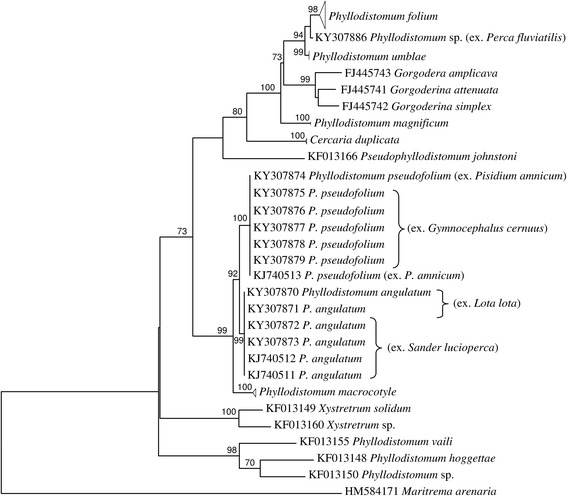
Results: Phylogenetic analyses of adult and larval stages of Phyllodistomum spp. based on ITS2 and partial 28S rDNA data allowed the confirmation of the validity of P. pseudofolium. A macrocercous cercaria, known as Phyllodistomum sp. from P. amnicum is genetically identical to adult P. pseudofolium. Phyllodistomum megalorchis obtained from its type-host, Lota lota L., showed no genetic differences from P. angulatum parasitizing Sander lucioperca L. In our analysis, P. pseudofolium, P. angulatum and P. macrocotyle formed a highly supported clade despite the fact that these species appear to be associated with distinct patterns of first intermediate host identity and cercarial morphology. Some morphological differences between gravid specimens of P. pseudofolium and P. angulatum were observed and their SEM tegumental surface topography is described.
Conclusions: The results lead us to the perception that macroevolutionary host switching in the genus Phyllodistomum is independent of host phylogeny. This study suggests strict host-specificity (oioxeny) for P. pseudofolium using one first intermediate host species (P. amnicum) and one definitive host species (G. cernuus). Phyllodistomum megalorchis is to be regarded as a synonym of P. angulatum. The close phylogenetic relatives, P. pseudofolium and P. angulatum, can be differentiated by morphological traits, the micromorphology and tegumental surface topography of these two species is intended to provide useful data for their identification and support the use of such features as a valuable taxonomic criterion. Molecular data showed that G. cernuus is a definitive host for two species: the oioxenous P. pseudofolium and the euryxenous P. folium.
Keywords: 28S; Eurasian ruffe; Host specificity; ITS2 rDNA; Life-cycles; Morphological variation; P. angulatum; Phyllodistomum pseudofolium; SEM.
Figures


Similar articles
-
Phyllodistomum kupermani n. sp. from the European perch, Perca fluviatilis L. (Perciformes: Percidae), and redescription of Phyllodistomum macrocotyle (Lühe, 1909) with notes on the species diversity and host specificity in the European Phyllodistomum spp. (Trematoda: Gorgoderidae).Parasit Vectors. 2020 Nov 10;13(1):561. doi: 10.1186/s13071-020-04434-2. Parasit Vectors. 2020. PMID: 33168101 Free PMC article.
-
Diversity and phylogenetic relationships of European species of Crepidostomum Braun, 1900 (Trematoda: Allocreadiidae) based on rDNA, with special reference to Crepidostomum oschmarini Zhokhov & Pugacheva, 1998.Parasit Vectors. 2018 Sep 28;11(1):530. doi: 10.1186/s13071-018-3095-y. Parasit Vectors. 2018. PMID: 30266086 Free PMC article.
-
Molecular and karyological identification and morphological description of cystocercous cercariae of Phyllodistomum umblae and Phyllodistomum folium (Digenea, Gorgoderidae) developing in European sphaeriid bivalves.Parasitol Int. 2015 Oct;64(5):441-7. doi: 10.1016/j.parint.2015.06.007. Epub 2015 Jun 24. Parasitol Int. 2015. PMID: 26116245
-
The use and implications of ribosomal DNA sequencing for the discrimination of digenean species.Adv Parasitol. 2005;60:101-63. doi: 10.1016/S0065-308X(05)60002-4. Adv Parasitol. 2005. PMID: 16230103 Review.
-
Review on the molecular study of the Diplozoidae: analyses of currently available genetic data, what it tells us, and where to go from here.Parasit Vectors. 2020 Oct 30;13(1):539. doi: 10.1186/s13071-020-04417-3. Parasit Vectors. 2020. PMID: 33126913 Free PMC article. Review.
Cited by
-
Intermediate insights: tracing trematodes infecting amphibians via their first intermediate snail hosts.Parasit Vectors. 2025 Jul 15;18(1):285. doi: 10.1186/s13071-025-06920-x. Parasit Vectors. 2025. PMID: 40665399 Free PMC article.
-
Cryptic speciation of the zoogonid digenean Diphterostomum flavum n. sp. demonstrated by morphological and molecular data.Parasite. 2020;27:44. doi: 10.1051/parasite/2020040. Epub 2020 Jun 18. Parasite. 2020. PMID: 32553099 Free PMC article.
-
Genetic characterization and phylogenetic relationships of Phyllodistomum parasites in Indian subcontinent: insights from freshwater fish and shrimp hosts.Parasitol Res. 2023 Oct;122(10):2301-2315. doi: 10.1007/s00436-023-07930-3. Epub 2023 Aug 23. Parasitol Res. 2023. PMID: 37610451 Free PMC article.
-
Amended diagnosis, validity and relationships of the genus Acrolichanus Ward, 1917 (Digenea: Allocreadiidae) based on the 28S rRNA gene, and observations on its lineage diversity.Syst Parasitol. 2020 Apr;97(2):143-156. doi: 10.1007/s11230-020-09901-z. Epub 2020 Feb 17. Syst Parasitol. 2020. PMID: 32065373
-
Plesiochorus irwinorum n. sp. (Trematoda: Gorgoderidae) from the urinary bladder of the hawksbill turtle, Eretmochelys imbricata (Testudines: Cheloniidae), off the east coast of Australia.Syst Parasitol. 2022 Aug;99(4):447-466. doi: 10.1007/s11230-022-10038-4. Epub 2022 Apr 23. Syst Parasitol. 2022. PMID: 35461430 Free PMC article.
References
-
- Combes C. Parasitism. The ecology and evolution of intimate interactions. Chicago and London: The University of Chicago Press; 2001.
-
- Kudinova MA. [On the revision of system of the trematode genus Phyllodistomum Braun, 1899 (Gorgoderidae).] In: Shulman SS, editor. Ecological Parasitology. Petrozavodsk: Karelian Research Center RAS; 1994. p. 96-112. (In Russian).
-
- Campbell RA. Family Gorgoderidae Looss, 1899. In: Bray RA, Gibson DI, Jones A, editors. Keys to the Trematoda, vol. 3. Wallingford: CABI Publishing and the Natural History Museum; 2008. p. 191–213.
MeSH terms
Substances
LinkOut - more resources
Full Text Sources
Other Literature Sources
Molecular Biology Databases
Miscellaneous
