Case Reports
. 2017 Jun 6;10(1):194.
doi: 10.1186/s13104-017-2513-0.

Pathohistological investigation of osteochondral tissue obtained during total knee arthroplasty after osteochondral autologous transfer: a case report

Affiliations
Case Reports

Pathohistological investigation of osteochondral tissue obtained during total knee arthroplasty after osteochondral autologous transfer: a case report

Momoko Tanima-Nagai et al. BMC Res Notes. .

Abstract

Background: Osteochondral autologous transfer is one of the repair techniques for cartilage defects of knee with promising knee function recovery. There are no reports including histopathological images concerning human osteochondral tissue after osteochondral autologous transfer. This is the first report to present pathohistological findings of transplanted plugs and host tissues extracted from the human body 3 years after osteochondral autologous transfer. This study aimed to explore the cause factor of chronic pain using histological techniques.

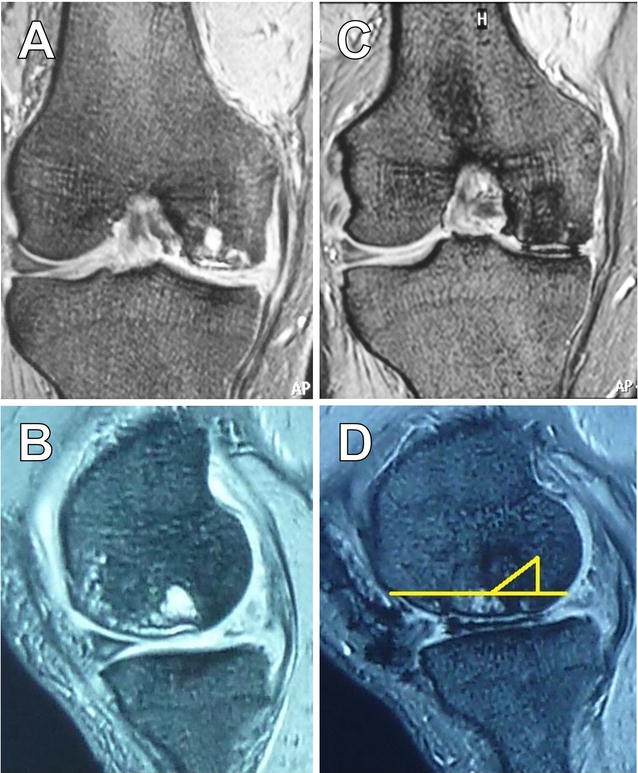
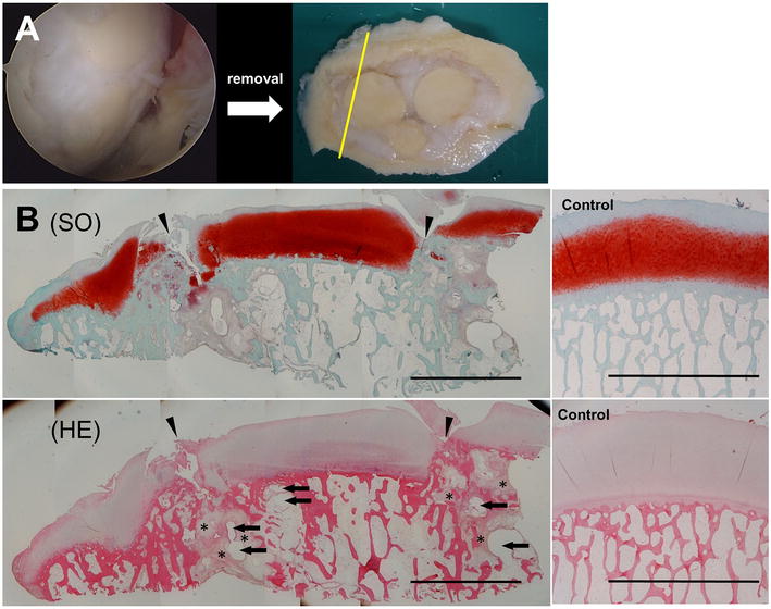
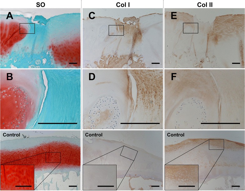
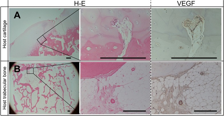
Case presentation: A 67-year-old Japanese man presented with adjusted total knee arthroplasty 3 years after osteochondral autologous transfer. Although in pain, arthroscopic assessment was not severe. The specimens which was gained during total knee arthroplasty were investigated in gross and microscopically using immunohistochemical staining technic. Histological examination revealed that the gap between grafted plugs and host osteochondral tissues was filled with fibrous tissue that stained positive for type I collagen. A degenerative change and some neovascularity were observed in the regenerated tissue and host trabecular bone. Furthermore, cysts and bone marrow edema were observed.

Conclusion: Our data suggests that the host osteochondral morbidity around grafted plugs might be related to chronical pain and revision surgery.

Keywords: Bone marrow edema; Case report; Osteochondral autologous transfer; Pathohistology.


Figures

Fig. 1
Fig. 1
Magnetic resonance image of pre- and post-osteochondral autologous transfer operation. Low intensity on T2-weighted images of the right medial femoral condyle (A, C coronal plane; B, D sagittal plane). The images were taken before osteochondral autologous transfer (A, B) and before total knee arthroplasty (C, D). Yellow lines show the resection line of two parts of the tissue
Fig. 2
Fig. 2
Arthroscopic images after osteochondral autologous transfer (OAT), and macro and micro observation images of isolated tissue after total knee arthroplasty (TKA). A At 1.5 years after OAT, good prognosis of the transferred grafts was confirmed by arthroscopy (left side). The osteochondral tissue contained inserted plugs that were discarded during TKA. The surface of the tissue was almost smooth (right side). The yellow line indicates the section area of (B). B Histological images with safranin-O staining (upper; SO) and hematoxylin–eosin staining (lower; H–E) show that the cartilage surface was not smooth between the host and plug cartilage. There were some cracks in the regenerated fibrous cartilage that reached bone marrow region. Arrowhead indicates the site of transferred plugs. Asterisk indicates bone marrow edema. Arrows indicate subchondral bone cyst. The control tissue shows the smooth surface of cartilage and strong staining intensity of SO. Scale bars indicate 5 mm
Fig. 3
Fig. 3
Histological and immunohistological stained images of repaired tissue between plugs and control tissue. Repair tissue between plugs with safranin-O (A, B), type I collagen (Col I; C, D), and type II collagen (Col II; E, F) show that the repaired tissue was not hyaline cartilage because plug cartilage and cluster cells existed in the cartilage adjacent to the repaired tissue. A, C, E Low magnification (×20). B, D, F High magnification images of quadrilateral area in above images (×200). Scale bars indicate 600 μm
Fig. 4
Fig. 4
The results of vascular endothelial growth factor (VEGF) expression and hematoxylin–eosin (H–E) staining at host cartilage and host trabecular bone. VEGF-positive cells and microvascular were often confirmed in the subchondral region connected to the cartilage region (A: upper lane) and in the host trabecular bone region (B: lower lane). Scale bars indicate 200 μm

Similar articles

References

    1. Hangody L, Vásárhelyi G, Hangody LR, Sükösd Z, Tibay G, Bartha L, Bodó G. Autologous osteochondral grafting–technique and long-term results. Injury. 2008;39(Suppl 1):S32–S39. doi: 10.1016/j.injury.2008.01.041. - DOI - PubMed
    1. Solheim E, Hegna J, Øyen J, Harlem T, Strand T. Results at 10 to 14 years after osteochondral autografting (mosaicplasty) in articular cartilage defects in the knee. Knee. 2013;20:287–290. doi: 10.1016/j.knee.2013.01.001. - DOI - PubMed
    1. Levy YD, Görtz S, Pulido PA, McCauley JC, Bugbee WD. Do fresh osteochondral allografts successfully treat femoral condyle lesions? Clin Orthop Relat Res. 2013;471:231–237. doi: 10.1007/s11999-012-2556-4. - DOI - PMC - PubMed
    1. De Caro F, Bisicchia S, Amendola A, Ding L. Large fresh osteochondral allografts of the knee: a systematic clinical and basic science review of the literature. Arthroscopy. 2015;31:757–765. doi: 10.1016/j.arthro.2014.11.025. - DOI - PubMed
    1. Lynch TS, Patel RM, Benedick A, Amin NH, Jones MH, Miniaci A. Systematic review of autogenous osteochondral transplant outcomes. Arthroscopy. 2015;31:746–754. doi: 10.1016/j.arthro.2014.11.018. - DOI - PubMed

Publication types

Substances