. 2017 Jun 6;17(1):96.
doi: 10.1186/s12903-017-0382-5.

Bactericidal effects of 310 nm ultraviolet light-emitting diode irradiation on oral bacteria

Affiliations

Bactericidal effects of 310 nm ultraviolet light-emitting diode irradiation on oral bacteria

Ayuko Takada et al. BMC Oral Health. .

Abstract

Background: Ultraviolet (UV) light is used for phototherapy in dermatology, and UVB light (around 310 nm) is effective for treatment of psoriasis and atopic dermatitis. In addition, it is known that UVC light (around 265 nm) has a bactericidal effect, but little is known about the bactericidal effect of UVB light. In this study, we examined the bactericidal effects of UVB-light emitting diode (LED) irradiation on oral bacteria to explore the possibility of using a 310 nm UVB-LED irradiation device for treatment of oral infectious diseases.

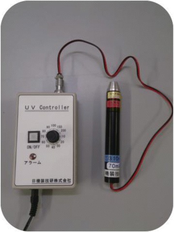
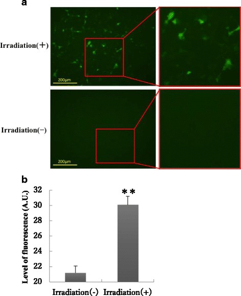
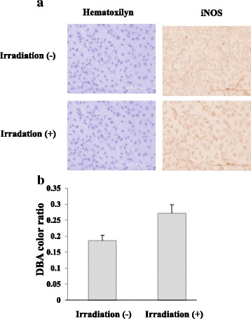
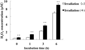
Methods: We prepared a UVB (310 nm) LED device for intraoral use to examine bactericidal effects on Streptococcus mutans, Streptococcus sauguinis, Porphyromonas gingivalis, and Fusobacterium nucleatum and also to examine the cytotoxicity to a human oral epithelial cell line (Ca9-22). We also examined the production of nitric oxide and hydrogen peroxide from Ca9-22 cells after irradiation with UVB-LED light.

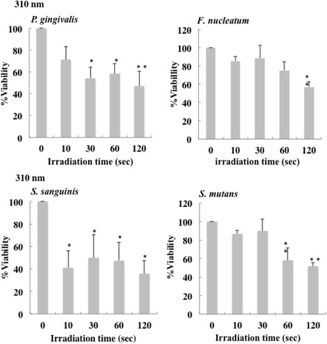
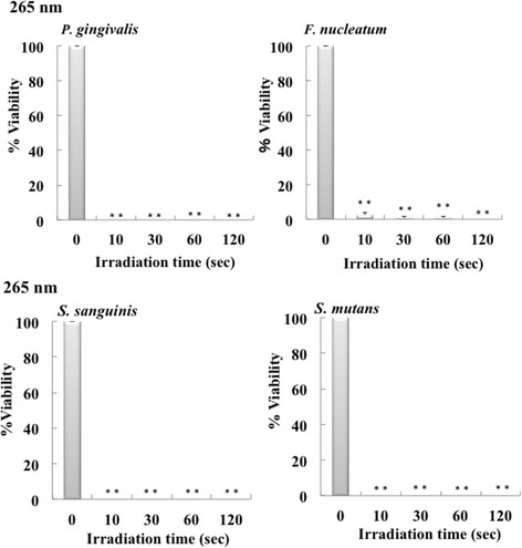
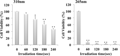
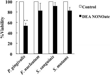
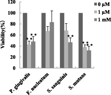
Results: Irradiation with the 310 nm UVB-LED at 105 mJ/cm2 showed 30-50% bactericidal activity to oral bacteria, though 17.1 mJ/cm2 irradiation with the 265 nm UVC-LED completely killed the bacteria. Ca9-22 cells were strongly injured by irradiation with the 265 nm UVC-LED but were not harmed by irradiation with the 310 nm UVB-LED. Nitric oxide and hydrogen peroxide were produced by Ca9-22 cells with irradiation using the 310 nm UVB-LED. P. gingivalis was killed by applying small amounts of those reactive oxygen species (ROS) in culture, but other bacteria showed low sensitivity to the ROS.

Conclusions: Narrowband UVB-LED irradiation exhibited a weak bactericidal effect on oral bacteria but showed low toxicity to gingival epithelial cells. Its irradiation also induces the production of ROS from oral epithelial cells and may enhance bactericidal activity to specific periodontopathic bacteria. It may be useful as a new adjunctive therapy for periodontitis.

Keywords: Periodontitis; Phototherapy; Porphyromonas gingivalis; Reactive oxygen species; Selective toxicity.


Figures

Fig. 1
Fig. 1
The prototype model of UVB (310 nm) and UVC (265 nm) LED irradiation device
Fig. 2
Fig. 2
Bactericidal effect of 310 nm UVB-LED irradiation on oral bacteria. Each bacterial suspension (0.1 ml) in 96-well plates was irradiated by a UV-LED. The suspensions were serially diluted and incubated on agar plates at 37 °C anaerobically. Colonies on the plates were counted after incubation for 1–7 days. Bactericidal levels are indicated as viability (%). (n = 3 or 5, means ± SE; **P < 0.01 vs. 0 s, *P < 0.05 vs. 0 s)
Fig. 3
Fig. 3
Bactericidal effect of 265 nm UV-LED irradiation on oral bacteria. Each of bacterial suspension (0.1 ml) in 96-well plates was irradiated by a UV-LED. The suspensions were serially diluted and incubated on agar plates at 37 °C anaerobically. Colonies on the plates were counted after incubation for 1–7 days. Bactericidal levels are indicated as viability (%). (n = 3, means ± SE; **P < 0.01 vs. 0 s)
Fig. 4
Fig. 4
Effect of 310 nm UVB-LED on human gingival epithelial cells. Ca9–22 cells were seeded at a density of 2 × 104 cells/well in 96-well cell culture plates. The cells were irradiated by a UV-LED (265 nm or 310 nm) for 60–240 s and were incubated for 24 h. Cytotoxicity of UV LED irradiation to the cells was measured by WST-8 assay. (n = 3, means ± SE; **P < 0.01 vs. 0 s,*P < 0.05 vs. 0 s)
Fig. 5
Fig. 5
Production of nitric oxide in Ca9–22 cells by irradiation with the 310 nm UVB-LED. a Monolayers of Ca9–22 cells were irradiated with 310 nm UVB-LED for 60 s and incubated for 24 h. Nitric oxide production into cells was then examined using DAF2-DA. b The level of fluorescence was expressed by using Image J. (n = 3, means ± SE; **P < 0.01)
Fig. 6
Fig. 6
Expression of iNOS in Ca9–22 cells by irradiation with the 310 nm UVB-LED. Monolayers of Ca9–22 cells in a chamber slide were irradiated by the 310 nm UVB-LED for 60 s and incubated for 24 h. a Expression of iNOS in the cells was measured by immunofluorescent staining using an anti-iNOS antibody. b The levels of DAB color ratio were measured by using Image J. (n = 3, means ± SE; P = 0.0503)
Fig. 7
Fig. 7
Production of hydrogen peroxide in Ca9–22 cells by irradiation with the 310 nm UVB-LED. Monolayers of Ca9–22 cells were irradiated with 310 nm UVB-LED for 60 s and incubated for 0–6 h. Hydrogen peroxide production into cells was then examined using a kit
Fig. 8
Fig. 8
Bactericidal effect of nitric oxide on oral bacteria. Each bacterial suspension (OD of 0.4–0.5 at 600 nm) was incubated with 10 μM DEA NONOate for 1 h. Serially diluted suspensions were seeded on agar plates and incubated for 1–10 days. Colonies on the plates were counted and viability of each bacterium was evaluated. (n = 3, means ± SE; **P < 0.01 vs. Control)
Fig. 9
Fig. 9
Bactericidal effect of hydrogen peroxide on oral bacteria. H2O2 (1 μM or 1 mM) was added to each 0.1 ml bacterial suspension (OD of 0.4–0.5 at 600 nm) in 96-well plates and the bacterial suspensions were incubated for 1 h. The serially diluted suspensions were seeded on agar plates, and CFUs were counted. (n = 3, means ± SE; **P < 0.01 vs. 0 μM, *P < 0.05 vs. 0 μM)

Similar articles

Cited by

References

    1. Lapolla W, Yentzer BA, Bagel J, Halvorson CR, Feldman SR. A review of phototherapy protocols for psoriasis treatment. J Am Acad Dermatol. 2011;64(5):936–949. doi: 10.1016/j.jaad.2009.12.054. - DOI - PubMed
    1. Patrizi A, Raone B, Ravaioli GM. Management of atopic dermatitis: safety and efficacy of phototherapy. Clin Cosmet Investig Dermatol. 2015;8:511–520. doi: 10.2147/CCID.S87987. - DOI - PMC - PubMed
    1. Shintani Y, Yasuda Y, Kobayashi K, Maeda A, Morita A. Narrowband ultraviolet B radiation suppresses contact hypersensitivity. Photodermatol Photoimmunol Photomed. 2008;24(1):32–37. doi: 10.1111/j.1600-0781.2008.00333.x. - DOI - PubMed
    1. Fernandez-Guarino M, Aboin-Gonzalez S, Barchino L, Velazquez D, Arsuaga C, Lazaro P. Treatment of moderate and severe adult chronic atopic dermatitis with narrow-band UVB and the combination of narrow-band UVB/UVA phototherapy. Dermatol Ther. 2016;29(1):19–23. doi: 10.1111/dth.12273. - DOI - PubMed
    1. Vangipuram R, Feldman SR. Ultraviolet phototherapy for cutaneous diseases: a concise review. Oral Dis. 2015;22(4):253–259. doi: 10.1111/odi.12366. - DOI - PubMed

MeSH terms