. 2017 Jun 6;36(1):76.
doi: 10.1186/s13046-017-0546-9.

In vitro antineoplastic effects of brivaracetam and lacosamide on human glioma cells

Affiliations

In vitro antineoplastic effects of brivaracetam and lacosamide on human glioma cells

Ambra Rizzo et al. J Exp Clin Cancer Res. .

Abstract

Background: Epilepsy is a frequent symptom in patients with glioma. Although treatment with antiepileptic drugs is generally effective in controlling seizures, drug-resistant patients are not uncommon. Multidrug resistance proteins (MRPs) and P-gp are over-represented in brain tissue of patients with drug-resistant epilepsy, suggesting their involvement in the clearance of antiepileptic medications. In addition to their anticonvulsant action, some drugs have been documented for cytotoxic effects. Aim of this study was to evaluate possible in vitro cytotoxic effects of two new-generation antiepileptic drugs on a human glioma cell line U87MG.

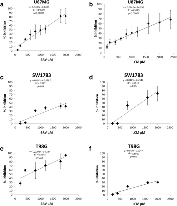
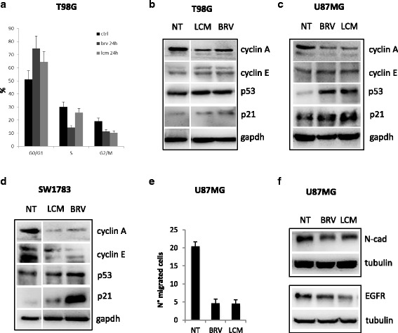
Methods: Cytotoxicity of brivaracetam and lacosamide was tested on U87MG, SW1783 and T98G by MTS assay. Expression of chemoresistance molecules was evaluated using flow cytometry in U87MG and human umbilical vein endothelial cells (HUVECs). To investigate the putative anti-proliferative effect, apoptosis assay, microRNA expression profile and study of cell cycle were performed.

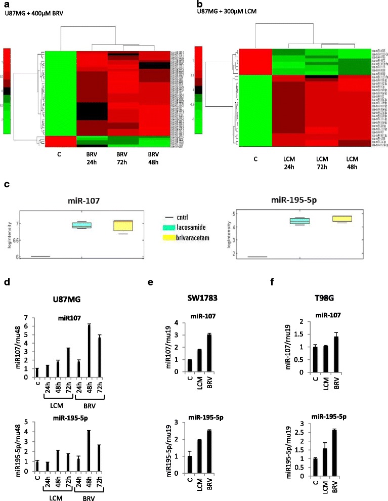
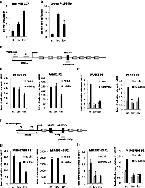
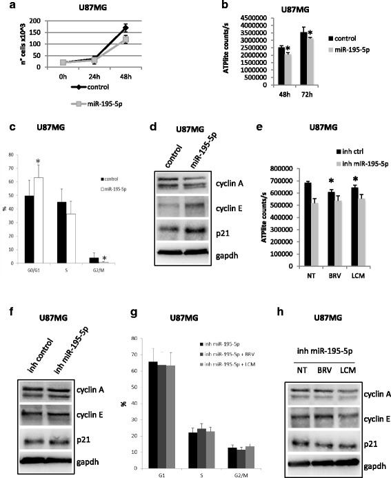
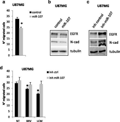
Results: Brivaracetam and lacosamide showed a dose-dependent cytotoxic and anti-migratory effects. Cytotoxicity was not related to apoptosis. The exposure of glioma cells to brivaracetam and lacosamide resulted in the modulation of several microRNAs; particularly, the effect of miR-195-5p modulation seemed to affect cell cycle, while miR-107 seemed to be implicated in the inhibition of cells migration. Moreover, brivaracetam and lacosamide treatment did not modulate the expression of chemoresistance-related molecules MRPs1-3-5, GSTπ, P-gp on U87MG and HUVECs.

Conclusion: Based on antineoplastic effect of brivaracetam and lacosamide on glioma cells, we assume that patients with glioma could benefit by the treatment with these two molecules, in addition to standard therapeutic options.

Keywords: Brain tumor-related epilepsy; Brivaracetam; Cytotoxicity; Glioma; Lacosamide.


Figures

Fig. 1
Fig. 1
Brivaracetam and lacosamide treatments exert a cytotoxic effect on U87MG, SW1783 and T98G glioma cells exposed to increasing drugs concentration. Graphs show the cytotoxic effect of BRV and LCM on U87MG cell line (a-b), Pearson correlation index p <0.00001 for both), SW1783 (c-d), Pearson correlation index p <0.05 for both) and T98G (e-f), Pearson correlation index p <0.05 for both). Data are expressed as % of inhibition calculated with the formula: 100-(100 x mean cell number x C/n.cell basal level) where C = drug concentration [range 0–2500 μM]. Data refer to at least three independent experiments, error bars represent the SD
Fig. 2
Fig. 2
Brivaracetam and lacosamide treatments exerted an anti-proliferative effect and a reduction in migration ability of glioma cells. a Distribution of T98G cells in the different phases of the cell cycle upon 24 h BRV or LCM treatments (IC20). Data are expressed as percentage of cell in a specific phase (G0/G1, S, G2/M) and refers to at least four independent experiments. Statistical evaluation was performed by the student’s t-test. Histogram bars represent mean ± standard deviation of at least three independent replicates. b-c-d Western-blot analysis of cyclin A, cyclin E, p53 and p21 upon treatments with BRV or LCM at IC20 in T98G a U87MG b SW1783 c cells. e Transwell migration assay in U87MG cells upon BRV or LCM treatments. (IC20). f Western-blot analysis of EGFR and N-cadherin proteins expression levels in U87MG cells upon BRV or LCM treatments (IC20)
Fig. 3
Fig. 3
Brivaracetam and lacosamide treatments modulate microRNAs expression in U87MG glioma cells. a Heat map of the identified signature of 37 microRNAs differentially expressed in U87MG cells treated with BRV at IC20 at the indicated time points. b Heat map of the identified signature of 30 microRNAs differentially expressed in U87MG cells treated with LCM at IC20 at the indicated time points. c Supervised statistical test of the significance level of the difference between signal distributions of miR-107 and miR-195-5p in U87MG cells treated with LCM or BRV versus the control (untreated cells). d-e-f qRT-PCR validation of miR-107 and miR-195-5p in U87MG, SW1783 and T98G upon IC20 LCM or BRV treatments at the indicated time points
Fig. 4
Fig. 4
Brivaracetam and lacosamide treatments induce epigenetic modification on miR-107 and miR-195 regulatory regions. a-b qRT-PCR of miR-107 a and miR-195-5p b precursors (pre-miRNAs) in U87MG cells upon IC20 LCM or BRV treatments. c Schematic representation of miR-107 gene locus: two different regions of host gene (PANK1) promoter have been analyzed in ChIP assays (P1 and P2). d-e ChIP analysis of acetylated histone H4 and methylated histone H3 occupancy on miR-107 regulatory regions in U87MG cells upon treatment with BRV or LCM at IC20. f Schematic representation of miR-195-5p gene locus: two different regions of host gene (MIR497HG) promoter have been analyzed in ChIP assays (P1 and P2). g-h ChIP analysis of acetylated histone H4 and methylated histone H3 occupancy on miR-195-5p regulatory regions in U87MG cells upon treatment with BRV or LCM at IC20
Fig. 5
Fig. 5
Brivaracetam and lacosamide treatments exert their anti-proliferative effect in part trough miR-195-5p. a-b Proliferation assay (a) and viability assay b in U87MG cells transfected with miR-195-5p mimic or control. Cells were collected and counted at the indicated time points. c Distribution of U87MG cells in the different phases of the cell cycle. Data are expressed as percentage of cell in a specific phase (G0/G1, S, G2/M) and refers to at least four independent experiments. Statistical evaluation was performed by the student’s t-test. Histogram bars represent mean ± standard deviation of at least three independent replicates. d Western-blot analysis of cyclin A, cyclin E and p21 protein expression levels in U87MG cells upon miR-195-5p over-expression. e Viability assay in U87MG cells transfected with miR-195-5p inhibitor or control and treated with BRV or LCM at IC20. f Western-blot analysis of cyclin A, cyclin E and p21 protein expression levels in U87MG cells upon miR-195-5p depletion with miR-195-5p inhibitor (inh miR-195-5p). g Distribution of U87MG cells in the different phases of the cell cycle upon miR-195 depletion and treatments wit BRV or LCM at IC20. Data are expressed as percentage of cell in a specific phase (G0/G1, S, G2/M) and refers to at least four independent experiments. Statistical evaluation was performed by the student’s t-test. Histogram bars represent mean ± standard deviation of at least three independent replicates. h Western-blot analysis of cyclin A, cyclin E and p21 protein expression levels in U87MG cells upon miR-195-5p depletion with miR-195-5p inhibitor (inh miR-195-5p) and treatments wit BRV or LCM at IC20. (* = pval < 0.05)
Fig. 6
Fig. 6
Brivaracetam and lacosamide treatments inhibit glioma cells migration in part trough miR-107. a Transwell migration assay in U87MG cells upon miR-107 exogenous expression (* = pval < 0.05). b-c Western-blot analysis of EGFR and N-cadherin proteins expression levels in U87MG cells upon miR-107 over-expression with miR-107 mimic (miR-107) (b) or depletion with miR-107 inhibitor (inh miR-107) (c). d Transwell migration assay in U87MG cells upon miR-107 depletion with miR-107 inhibitor (inh miR-107)

Similar articles

Cited by

References

    1. Blomqvist P, Lycke J, Strang P, Tornqvist H, Ekbom A. Brain tumours in Sweden 1996: care and costs. J Neurol Neurosurg Psychiatry. 2000;69(6):792–8. doi: 10.1136/jnnp.69.6.792. - DOI - PMC - PubMed
    1. Ekman M, Westphal M. Cost of brain tumour in Europe. Eur J Neurol. 2005;12(Suppl 1):45–9. doi: 10.1111/j.1468-1331.2005.01189.x. - DOI - PubMed
    1. Gustavsson A, Svensson M, Jacobi F, Allgulander C, Alonso J, Beghi E, Dodel R, Ekman M, Faravelli C, Fratiglioni L, Gannon B, Jones DH, Jennum P, Jordanova A, Jonsson L, Karampampa K, et al. Cost of disorders of the brain in Europe 2010. Eur Neuropsychopharmacol. 2011;21(10):718–79. doi: 10.1016/j.euroneuro.2011.08.008. - DOI - PubMed
    1. Latif AZ, Signorini D, Gregor A, Whittle IR. The costs of managing patients with malignant glioma at a neuro-oncology clinic. Br J Neurosurg. 1998;12(2):118–22. doi: 10.1080/02688699845230. - DOI - PubMed
    1. Maschio M, Sperati F, Dinapoli L, Vidiri A, Fabi A, Pace A, Pompili A, Carapella CM, Cantelmi T. Weight of epilepsy in brain tumor patients. J Neuro-Oncol. 2014;118(2):385–93. doi: 10.1007/s11060-014-1449-7. - DOI - PubMed

LinkOut - more resources