Review
. 2017 Nov 10;429(22):3409-3429.
doi: 10.1016/j.jmb.2017.05.029. Epub 2017 Jun 3.

Molecular Basis for K63-Linked Ubiquitination Processes in Double-Strand DNA Break Repair: A Focus on Kinetics and Dynamics

Affiliations
Review

Molecular Basis for K63-Linked Ubiquitination Processes in Double-Strand DNA Break Repair: A Focus on Kinetics and Dynamics

Brian L Lee et al. J Mol Biol. .

Abstract

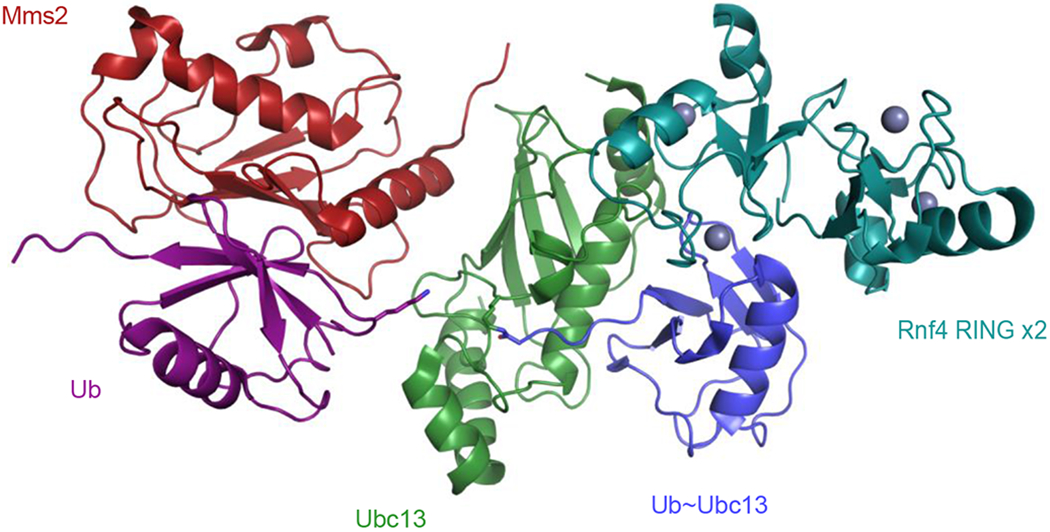
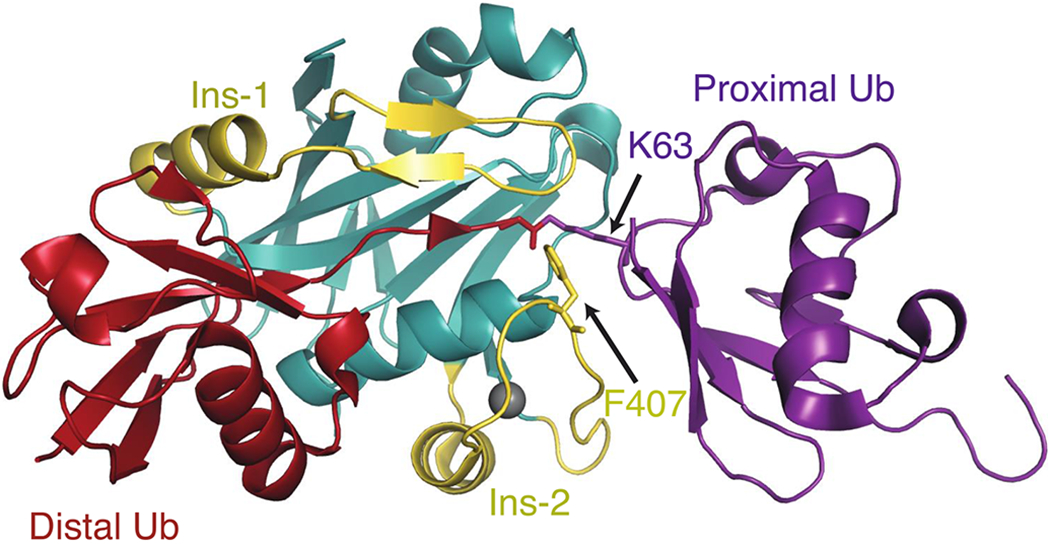
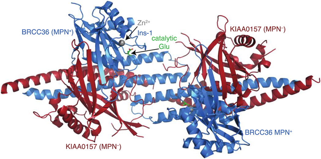
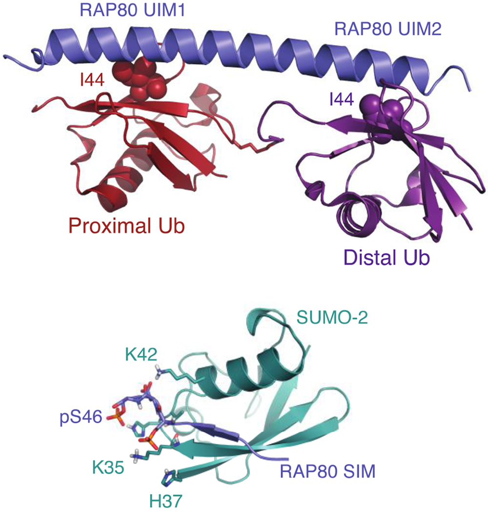
Cells are exposed to thousands of DNA damage events on a daily basis. This damage must be repaired to preserve genetic information and prevent development of disease. The most deleterious damage is a double-strand break (DSB), which is detected and repaired by mechanisms known as non-homologous end-joining (NHEJ) and homologous recombination (HR), which are components of the DNA damage response system. NHEJ is an error-prone first line of defense, whereas HR invokes error-free repair and is the focus of this review. The functions of the protein components of HR-driven DNA repair are regulated by the coordinated action of post-translational modifications including lysine acetylation, phosphorylation, ubiquitination, and SUMOylation. The latter two mechanisms are fundamental for recognition of DSBs and reorganizing chromatin to facilitate repair. We focus on the structures and molecular mechanisms for the protein components underlying synthesis, recognition, and cleavage of K63-linked ubiquitin chains, which are abundant at damage sites and obligatory for DSB repair. The forward flux of the K63-linked ubiquitination cascade is driven by the combined activity of E1 enzyme, the heterodimeric E2 Mms2-Ubc13, and its cognate E3 ligases RNF8 and RNF168, which is balanced through the binding and cleavage of chains by the deubiquitinase BRCC36, and the proteasome, and through the binding of chains by recognition modules on repair proteins such as RAP80. We highlight a number of aspects regarding our current understanding for the role of kinetics and dynamics in determining the function of the enzymes and chain recognition modules that drive K63 ubiquitination.

Keywords: DNA damage response; enzyme kinetics; protein dynamics; protein–protein-interactions; ubiquitination.


Figures

Figure 1.
Figure 1.
Structure of ubiquitin (blue) attached to Ubc13 (green) through a stable isopeptide linkage representing Ub~Ubc13 thioester (sticks), interacting with the RNF4 RING domain (cyan), and Mms2 (red), which orients a proximal (acceptor) Ub (purple) K63 sidechain (sticks) for nucleophilic attack of the thioester bond.
Figure 2.
Figure 2.
Structure of AMSH-LP bound to K63-Ub2 (2ZNV) showing the JAMM, or MPN+ domain (turquoise) and associated Ins-1 and -2 loops (yellow), the distal (donor, red) and proximal (acceptor, purple) Ubs, respectively. The C-terminal distal Gly, acceptor K63, and F407 at the apex of the flexible Ins-2 loop are shown in the stick representation.
Figure 3.
Figure 3.
Structure of the BRCC36-KIAA0157 MPN+–MPN heterodimeric super dimer (5CW3), with BRCC36 (blue), KIAA0157 (red) in the ribbon representation with Ins-1 and the catalytic Zn2+, and active site Ins-1 loop and Glu (green, stick representation) indicated.
Figure 4.
Figure 4.
Structure of the RAP80 tandem UIMs (blue) bound to K63-Ub2 (top panel), showing the proximal (acceptor, red), and distal (donor, purple), with Ile44 at the center of the Ub hydrophobic patch in the van der Waals representation, and with acceptor K63 and donor C-terminal Gly in the stick representation. The RAP80 SIM (blue) interaction with SUMO-2 (turquoise) is shown in the bottom panel. Positively charged SUMO-2 residues that interact with negatively charged phosphates from the RAP80 SIM are indicated, and shown in the stick representation.

Similar articles

Cited by

References

    1. Jackson SP, Bartek J, The DNA-damage response in human biology and disease, Nature. 461 (2009) 1071–1078. doi:10.1038/nature08467. - DOI - PMC - PubMed
    1. Kastan MB, Bartek J, Cell-cycle checkpoints and cancer, Nature. 432 (2004) 316–323. doi:10.1038/nature03097. - DOI - PubMed
    1. Ciccia A, Elledge SJ, The DNA damage response: Making it safe to play with knives, Mol. Cell 40 (2010) 179–204. doi:10.1016/j.molcel.2010.09.019. - DOI - PMC - PubMed
    1. Schwertman P, Bekker-Jensen S, Mailand N, Regulation of DNA double-strand break repair by ubiquitin and ubiquitin-like modifiers, Nat. Rev. Mol. Cell Biol 17 (2016) 379–394. doi:10.1038/nrm.2016.58. - DOI - PubMed
    1. Bekker-Jensen S, Mailand N, Assembly and function of DNA double-strand break repair foci in mammalian cells, DNA Repair. 9 (2010) 1219–1228. doi:10.1016/j.dnarep.2010.09.010. - DOI - PubMed

Publication types

LinkOut - more resources