. 2017 Jul 3;214(7):1973-1989.
doi: 10.1084/jem.20170495. Epub 2017 Jun 6.

Commensal microbes provide first line defense against Listeria monocytogenes infection

Affiliations

Commensal microbes provide first line defense against Listeria monocytogenes infection

Simone Becattini et al. J Exp Med. .

Abstract

Listeria monocytogenes is a foodborne pathogen that causes septicemia, meningitis and chorioamnionitis and is associated with high mortality. Immunocompetent humans and animals, however, can tolerate high doses of L. monocytogenes without developing systemic disease. The intestinal microbiota provides colonization resistance against many orally acquired pathogens, and antibiotic-mediated depletion of the microbiota reduces host resistance to infection. Here we show that a diverse microbiota markedly reduces Listeria monocytogenes colonization of the gut lumen and prevents systemic dissemination. Antibiotic administration to mice before low dose oral inoculation increases L. monocytogenes growth in the intestine. In immunodeficient or chemotherapy-treated mice, the intestinal microbiota provides nonredundant defense against lethal, disseminated infection. We have assembled a consortium of commensal bacteria belonging to the Clostridiales order, which exerts in vitro antilisterial activity and confers in vivo resistance upon transfer into germ free mice. Thus, we demonstrate a defensive role of the gut microbiota against Listeria monocytogenes infection and identify intestinal commensal species that, by enhancing resistance against this pathogen, represent potential probiotics.


Figures

Figure 1.
Figure 1.
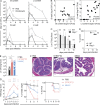
Antibiotic treatment predisposes to severe L. monocytogenes infection. (A) L. monocytogenes (Lm) burden in antibiotic-treated mice. WT mice were treated with a single i.p. injection of clindamycin or PBS and infected orally 24 h later with 107 Lm 10403s CFUs. At each time point, animals were euthanized and the total number of Lm CFUs was determined by plating homogenized organs or intestinal content (n = 4 per time point, from two independent experiments). (B) Spearman correlation between Lm CFUs recovered from the intestinal content and wall of mice shown in A, for small and large intestine. (C and D) WT and Rag1−/− (Rag) mice were cohoused for 3 wk, then injected i.p. with a single dose of clindamycin and infected 24 h later with 107 Lm CFUs. Survival (C) and fecal shedding (D) of Lm were monitored over time (geometric means + geometric SD are shown; n = 10 per group; from two independent experiments). (E) Lm burden in feces 1 d after infection with 108 Lm CFUs of mice treated with either streptomycin or a combination of metronidazole, neomycin, vancomycin, and clindamycin (MNVC). Antibiotic treatment was terminated 1 d before infection in both cases (n = 9–13; Kruskal-Wallis test with Dunn’s multiple comparison correction). (F) Representative H&E staining of colonic tissue from mice treated as in E, 3 d after infection (arrows indicate edema and stars indicate cellular infiltration; Bar, 200 µm). (G) Weight loss, pathology score (see Materials and methods) and survival of mice treated as in E (n = 13–17 per group; means + SD; Two way ANOVA with Tukey’s multiple comparison test and Log-Rank (Mantel-Cox) for survival). *, P < 0.05; **, P < 0.005; ***, P < 0.001; ****, P < 0.0001.
Figure 2.
Figure 2.
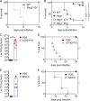
Small L. monocytogenes oral inocula spread systemically in antibiotic-treated mice. (A) WT mice were treated with one dose of oral streptomycin or PBS and infected 24 h later with 104 or 102 (only streptomycin group) CFUs of L. monocytogenes (Lm). Lm fecal shedding is shown over time (mean + SD; n = 8 per group; from two independent experiments). (B) Weight loss for animals shown in A (n = 7–8; n = 3 for streptomycin-only group; from two independent experiments; two Way ANOVA with Tukey’s multiple comparisons; a comma separates significance values for STREPTO × 104 vs. PBS × 104 groups and STREPTO × 102 vs. PBS × 104 groups, respectively). (C) Lm burden in the depicted compartments at 3 d after infection, same conditions as in A (n = 6; from two independent experiments; Kruskal-Wallis test with Dunn’s multiple comparisons). (D) Mice infected with 108 Lm particles were euthanized 1 d after negative fecal cultures for Lm, and whole organs/intestinal contents were homogenized and plated for Lm detection (n = 6; shown are only mice for which colonies where detected). (E) Kinetics of Lm fecal shedding in mice infected with 108 Lm CFUs. Lm presence in the feces was monitored over time, and mice were administered 1 dose of streptomycin (salmon arrow) on the first day after fecal cultures became negative for Lm (n = 4; one representative of three experiments shown). (F) Percentages of mice bearing Lm at the depicted time points after infection, as assessed using the approach described in E, except that in this experiment, mice were maintained in wire floor cages to prevent coprophagy (n = 10 per group; from three different experiments). *, P < 0.05; **, P < 0.005; ***, P < 0.001; ****, P < 0.0001.
Figure 3.
Figure 3.
Antibiotic treatment predisposes hosts with congenic immunodeficiency to lethal listeriosis. (A) Rag2−/−Il2rg−/− (Raggc) and WT mice were cohoused for 3 wk and infected orally with 108 L. monocytogenes (Lm) CFUs. Survival was monitored overtime (n = 3; similar results were obtained with a lower infectious dose). (B) Mice of the depicted strain were cohoused for 3 wk, and then challenged orally with 108 Lm CFUs. Survival was monitored overtime (n = 9–15; from three independent experiments). (C–F) Raggc mice were administered the depicted antibiotics or PBS and infected 24 h later with 104 Lm CFUs. (C and F) Show Lm CFUs in feces 1 d after infection (mean + SD), (D and F) show survival rates (n = 6–7 for [A and B]; n = 4 for [C and D], from three and two independent experiments, respectively; Mann-Withney test for CFU comparison and Log-Rank [Mantel-Cox] test for survival.) *, P < 0.05; **, P < 0.01; ***, P < 0.001; ****, P < 0.0001.
Figure 4.
Figure 4.
Anticancer chemotherapy and antibiotics synergistically enhance susceptibility to L. monocytogenes infection. (A) Mice were administered combined chemotherapy (CHEMO) composed of cyclophosphamide and doxorubicin, or PBS, injected twice i.p. (on day 0 and 7). On day 8 mice were bled and the white blood cell (WBC) count was determined (n = 14; from three independent experiments). (B) Representative FACS plots of cells obtained as in A and stained for markers of interest. (C) Cell numbers for circulating leukocytes identified as in B from mice shown in A (n = 14; lines represent means). (D) Survival of PBS- vs. CHEMO-treated mice infected 1 d after second treatment (d8) with 106 L. monocytogenes CFUs (n = 5). (E) Lm burden in the colonic content of mice treated and infected as in D with 108 Lm CFUs, 1 d after infection. Shown is one representative of two experiments (n = 4). (F) Mice were treated as in A, administered either PBS or streptomycin on day 7 (concomitant with the second CHEMO administration) and infected 24 h later with 104 Lm CFUs (n = 12–13; Mann-Whitney test in A, C, and E; Log-Rank test in D and F. *, P < 0.05; **, P < 0.005; ****, P < 0.0001).
Figure 5.
Figure 5.
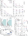
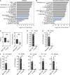
Intestinal microbes efficiently eliminate L. monocytogenes ex vivo, through diverse mechanisms. (A) Content from small and large intestine was collected from WT mice and resuspended in reduced PBS. L. monocytogenes (Lm) was inoculated at the depicted doses in 100 µl of intestinal suspension and cultured in anaerobic (top) or aerobic (bottom) conditions. Lm CFUs were enumerated over time by plating (n = 3 mice per time point; one representative of two experiments shown, circles represent individual values, lines represent medians). (B) 103 Lm CFUs were inoculated in intestinal contents prepared as in A or in sterile filtered aliquots of the same intestinal contents and grown anaerobically or aerobically, respectively, for the depicted times (n = 3 mice per time point; one representative of two experiments shown, lines represent medians). (C) Same experimental setup as in A, except that intestinal content was collected from PBS, streptomycin or MNVC-treated mice 1 d after termination of treatment (n = 3 mice per time point; one representative of two experiments shown, circles represent individual values, lines represent medians). (D) Content from small or large intestine was cultured for 24 h in the presence of 103 Lm CFUs, then sterile filtered (24 h-sup). 103 CFUs of either L. monocytogenes or E. coli DH5-α were inoculated in sterile-filtered supernatants (24 h-sup) and grown aerobically for 24 h (n = 10 from different mice and three independent experiments, lines represents medians; statistics: Kruskal-Wallis test with Dunn’s multiple comparisons). ***, P < 0.001; ****, P < 0.0001.
Figure 6.
Figure 6.
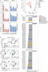
Correlation of intestinal commensal bacteria with protection from L. monocytogenes infection. (A) WT mice were treated with streptomycin or MNVC. Three mice per group were single-housed and infected with 108 Lm CFUs at each of the depicted time points (after antibiotic treatment). 24 h after infection, mice were sacrificed and L. monocytogenes CFUs enumerated in their intestinal content and organs. (B) Spearman correlation between Lm CFUs (Log10) in intestinal content, intestinal wall and liver for animals depicted in A (n = 30). Spearman coefficient and significance values are indicated for each correlation. (C) PCoA analysis of microbiota 16S sequences from fecal pellets collected from animals depicted in A on the day of infection. The plotted colored areas indicate: gray = pretreatment; green = d1 after antibiotics (any antibiotics); salmon = d5-27 streptomycin; blue = d5-27 MNVC. (D) Spearman correlation between identified OTUs and Lm CFUs enumerated by plating 1 d after infection, shown separately for small intestine, cecum, and colon contents. Shown are only significant hits, BH corrected. P < 0.05.
Figure 7.
Figure 7.
A rationally designed consortium of four Clostridiales antagonizes L. monocytogenes and confers host protection in vivo. (A and B) Depicted commensal bacteria were grown anaerobically and inoculated in medium (A) or in autoclaved intestinal content (B) at OD = 0.1. 103 L. monocytogenes (Lm) CFUs were added, and Lm expansion was evaluated after 24 h of anaerobic co-culture (n = 3 replicates; shown one representative of two to four experiments per condition). (C–E) GF mice were reconstituted with 4-Clost consortium or fecal pellet from MNVC-treated mice via oral gavage. After 10 d mice were challenged orally with 107 Lm CFUs. (C) Diversity index of the microbiota 10 d after reconstitution (=day of L. monocytogenes infection) in feces of ex-GF mice of the indicated group, based on OTU composition as assessed by sequencing of 16S rRNA genes. (D) Listeria burden in the feces of ex-GF animals was evaluated 24 h after infection. (E and F) Mice described in C and D were euthanized at day 3 after infection and L. monocytogenes CFUs quantified in intestinal content and depicted organs. For C–E, circles represent individual mice, bars represent median values (n = 9 except in C; n = 5, from three independent experiments; Mann-Whitney test). *, P < 0.05; **, P < 0.01; ***, P < 0.001; ****, P < 0.0001.

References

    1. Abt M.C., Lewis B.B., Caballero S., Xiong H., Carter R.A., Sušac B., Ling L., Leiner I., and Pamer E.G.. 2015. Innate Immune Defenses Mediated by Two ILC Subsets Are Critical for Protection against Acute Clostridium difficile Infection. Cell Host Microbe. 18:27–37. 10.1016/j.chom.2015.06.011 - DOI - PMC - PubMed
    1. Allerberger F., and Wagner M.. 2010. Listeriosis: a resurgent foodborne infection. Clin. Microbiol. Infect. 16:16–23. 10.1111/j.1469-0691.2009.03109.x - DOI - PubMed
    1. Alyamkina E.A., Nikolin V.P., Popova N.A., Dolgova E.V., Proskurina A.S., Orishchenko K.E., Efremov Y.R., Chernykh E.R., Ostanin A.A., Sidorov S.V., et al. . 2010. A strategy of tumor treatment in mice with doxorubicin-cyclophosphamide combination based on dendritic cell activation by human double-stranded DNA preparation. Genet. Vaccines Ther. 8:7 10.1186/1479-0556-8-7 - DOI - PMC - PubMed
    1. Andersson A., Dai W.J., Di Santo J.P., and Brombacher F.. 1998. Early IFN-gamma production and innate immunity during Listeria monocytogenes infection in the absence of NK cells. J. Immunol. 161:5600–5606. - PubMed
    1. Aoki S.K., Pamma R., Hernday A.D., Bickham J.E., Braaten B.A., and Low D.A.. 2005. Contact-dependent inhibition of growth in Escherichia coli. Science. 309:1245–1248. 10.1126/science.1115109 - DOI - PubMed

MeSH terms

Substances