

Diagnostic Performance of 18F-FDG PET/CT in Papillary Thyroid Carcinoma with Negative 131I-WBS at first Postablation, Negative Tg and Progressively Increased TgAb Level
- PMID: 28588229
- PMCID: PMC5460229
- DOI: 10.1038/s41598-017-03001-7
Diagnostic Performance of 18F-FDG PET/CT in Papillary Thyroid Carcinoma with Negative 131I-WBS at first Postablation, Negative Tg and Progressively Increased TgAb Level
Abstract
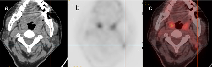
Differentiated thyroid cancer (DTC) patients with negative serum thyroglobulin (Tg), negative 131I whole-body scintigraphy (131I-WBS) at first post-ablation and progressively increased TgAb level are a relatively rare entity in the follow-up after total thyroidectomy and radioactive iodine therapy. The value of 18F-FDG PET/CT in detecting the recurrence of disease in these patients has only been reported in a small case series. The goal of this study was to investigate the diagnostic accuracy of 18F-FDG PET/CT in detecting recurrent disease in these specific PTC patients and to identify risk factors for patients with positive 18F-FDG PET/CT results. Eighty-two PTC patients who had 18F-FDG PET/CT scans with negative Tg, negative 131I-WBS at first post-ablation and progressively increased TgAb levels were included. We found that the sensitivity, specificity, positive predictive value, negative predictive value, and accuracy of 18F-FDG PET/CT in this patient group were determined as 84%, 72%, 92%, 57% and 82%, respectively. 18F-FDG PET/CT scan had a good diagnostic performance and should be performed routinely in PTC patients with negative Tg, negative 131I-WBS at first postablation and progressively increased TgAb level, especially when span for progressively increased TgAb level ≥ 3 years and/or progressively increased TgAb value up to 150 IU/mL.
Conflict of interest statement
The authors declare that they have no competing interests.
Figures

References
-
- Brierley J, Tsang R, Panzarella T, Bana N. Prognostic factors and the effect of treatment with radioactive iodine and external beam radiation on patients with differentiated thyroid cancer seen at a single institution over 40 years. Clin Endocrinol (Oxf) 2005;63:418–427. doi: 10.1111/j.1365-2265.2005.02358.x. - DOI - PubMed
-
- Filesi M, Signore A, Ventroni G, Melacrinis FF, Ronga G. Role of initial iodine-131 whole-body scan and serum thyroglobulin in differentiated thyroid carcinoma metastases. J Nucl Med. 1998;39:1542–1546. - PubMed
MeSH terms
Substances
LinkOut - more resources
Full Text Sources
Other Literature Sources
Miscellaneous
