

Single-axon level morphological analysis of corticofugal projection neurons in mouse barrel field
- PMID: 28588276
- PMCID: PMC5460143
- DOI: 10.1038/s41598-017-03000-8
Single-axon level morphological analysis of corticofugal projection neurons in mouse barrel field
Abstract
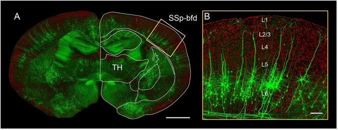
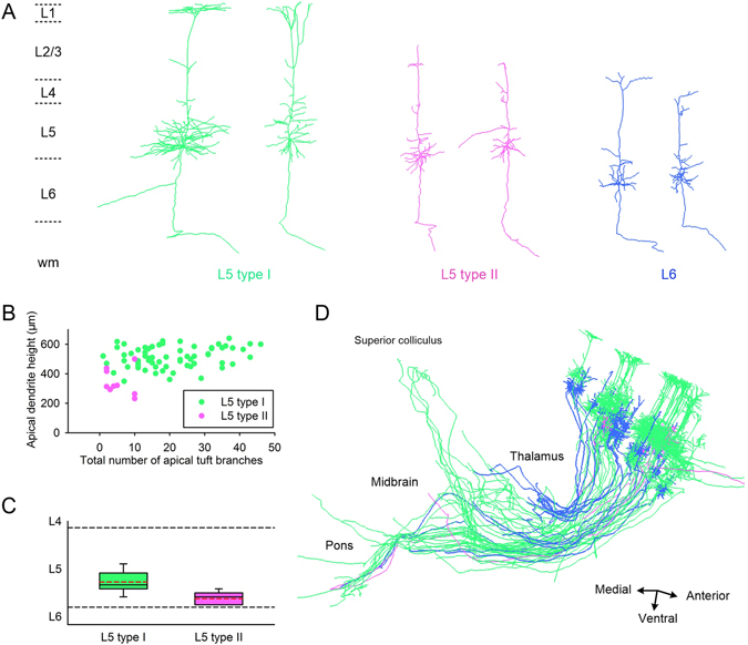
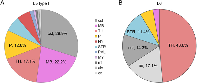
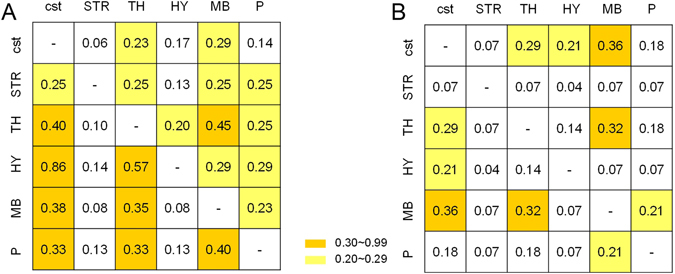
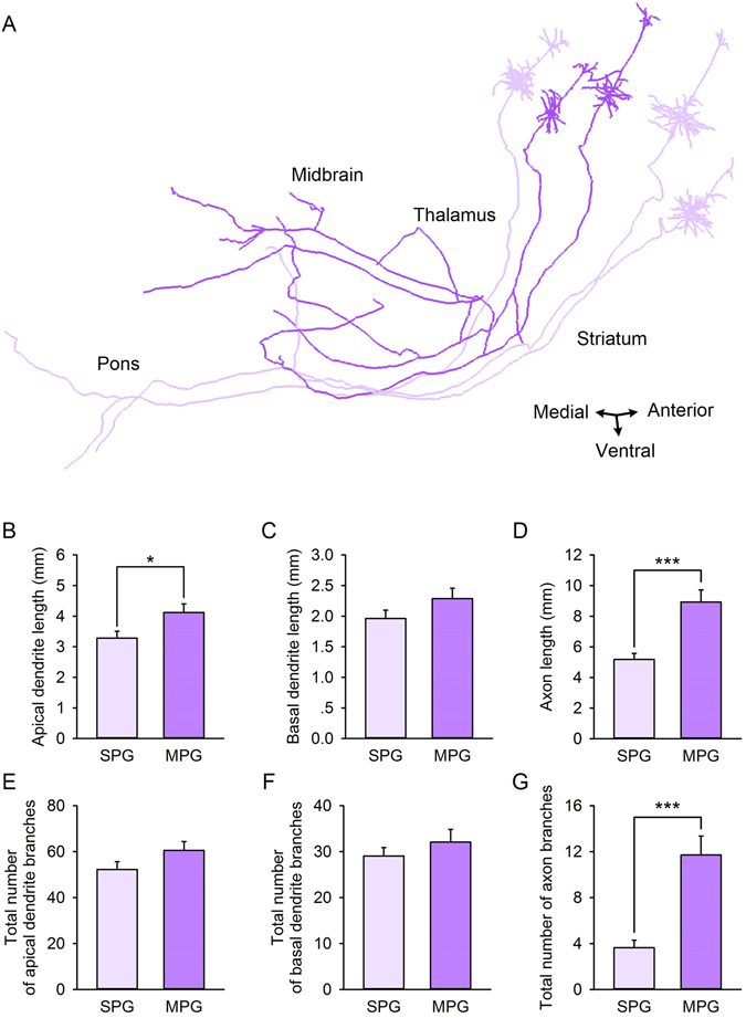
Corticofugal projection neurons are key components in connecting the neocortex and the subcortical regions. In the barrel field, these neurons have various projection targets and play crucial roles in the rodent whisker sensorimotor system. However, the projection features of corticofugal projection neurons at the single-axon level are far from comprehensive elucidation. Based on a brain-wide positioning system with high-resolution imaging for Thy1-GFP M-line mice brains, we reconstructed and analyzed more than one hundred corticofugal projection neurons in both layer V and VI of barrel cortex. The dual-color imaging made it possible to locate the neurons' somata, trace their corresponding dendrites and axons and then distinguish the neurons as L5 type I/II or L6 type. The corticofugal projection pattern showed significant diversity across individual neurons. Usually, the L5 type I neurons have greater multi-region projection potential. The thalamus and the midbrain are the most frequent projection targets among the investigated multidirectional projection neurons, and the hypothalamus is particularly unique in that it only appears in multidirectional projection situations. Statistically, the average branch length of apical dendrites in multi-region projection groups is larger than that of single-region projection groups. This study demonstrated a single-axon-level analysis for barrel corticofugal projection neurons, which could provide a micro-anatomical basis for interpreting whisker sensorimotor circuit function.
Conflict of interest statement
The authors declare that they have no competing interests.
Figures
Similar articles
-
Pinpointing Morphology and Projection of Excitatory Neurons in Mouse Visual Cortex.Front Neurosci. 2019 Aug 29;13:912. doi: 10.3389/fnins.2019.00912. eCollection 2019. Front Neurosci. 2019. PMID: 31555081 Free PMC article.
-
Anatomic and molecular development of corticostriatal projection neurons in mice.Cereb Cortex. 2014 Feb;24(2):293-303. doi: 10.1093/cercor/bhs342. Epub 2012 Oct 31. Cereb Cortex. 2014. PMID: 23118198 Free PMC article.
-
Epigenomic diversity of cortical projection neurons in the mouse brain.Nature. 2021 Oct;598(7879):167-173. doi: 10.1038/s41586-021-03223-w. Epub 2021 Oct 6. Nature. 2021. PMID: 34616065 Free PMC article.
-
Re: Woolsey TA, van der Loos H. 1970. The structural organization of layer IV in the somatosensory region (S I) of mouse cerebral cortex. Brain Res. 17: 205-242.Brain Res. 2016 Aug 15;1645:22-4. doi: 10.1016/j.brainres.2016.04.029. Epub 2016 Apr 14. Brain Res. 2016. PMID: 27086973 Review.
-
Neocortical layer 5 subclasses: From cellular properties to roles in behavior.Front Synaptic Neurosci. 2022 Oct 28;14:1006773. doi: 10.3389/fnsyn.2022.1006773. eCollection 2022. Front Synaptic Neurosci. 2022. PMID: 36387773 Free PMC article. Review.
Cited by
-
Cortical Output Is Gated by Horizontally Projecting Neurons in the Deep Layers.Neuron. 2020 Jan 8;105(1):122-137.e8. doi: 10.1016/j.neuron.2019.10.011. Epub 2019 Nov 26. Neuron. 2020. PMID: 31784285 Free PMC article.
-
Corticothalamic Pathways From Layer 5: Emerging Roles in Computation and Pathology.Front Neural Circuits. 2021 Sep 9;15:730211. doi: 10.3389/fncir.2021.730211. eCollection 2021. Front Neural Circuits. 2021. PMID: 34566583 Free PMC article. Review.
-
Acetylcholine deficiency disrupts extratelencephalic projection neurons in the prefrontal cortex in a mouse model of Alzheimer's disease.Nat Commun. 2022 Feb 22;13(1):998. doi: 10.1038/s41467-022-28493-4. Nat Commun. 2022. PMID: 35194025 Free PMC article.
-
High-Throughput Mapping of Long-Range Neuronal Projection Using In Situ Sequencing.Cell. 2019 Oct 17;179(3):772-786.e19. doi: 10.1016/j.cell.2019.09.023. Cell. 2019. PMID: 31626774 Free PMC article.
-
Automated Brain Region Segmentation for Single Cell Resolution Histological Images Based on Markov Random Field.Neuroinformatics. 2020 Apr;18(2):181-197. doi: 10.1007/s12021-019-09432-z. Neuroinformatics. 2020. PMID: 31376002
References
Publication types
MeSH terms
Substances
LinkOut - more resources
Full Text Sources
Other Literature Sources
Miscellaneous
