. 2017 Jun 6;7(1):2835.
doi: 10.1038/s41598-017-03013-3.

A Central Small Amino Acid in the VAMP2 Transmembrane Domain Regulates the Fusion Pore in Exocytosis

Affiliations

A Central Small Amino Acid in the VAMP2 Transmembrane Domain Regulates the Fusion Pore in Exocytosis

Benoît Hastoy et al. Sci Rep. .

Abstract

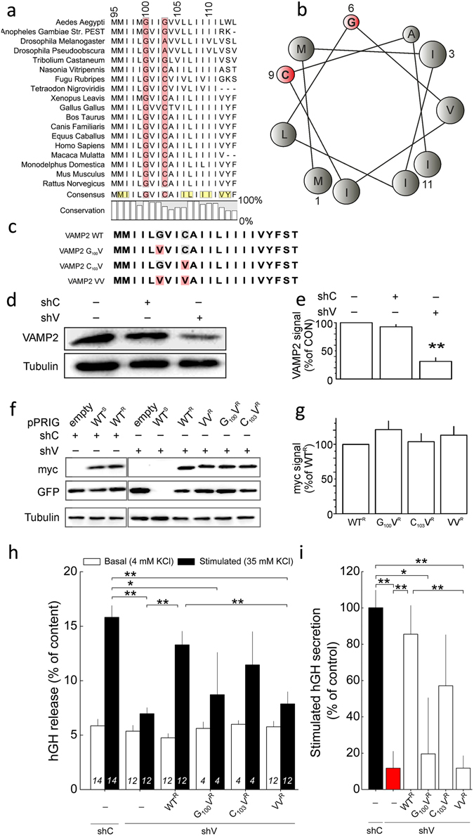
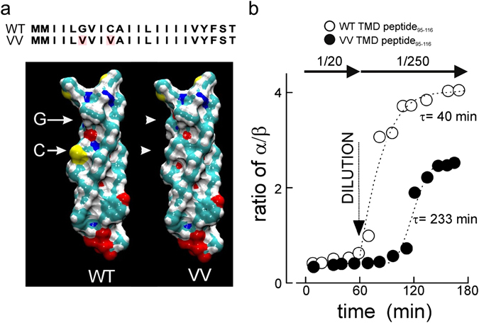
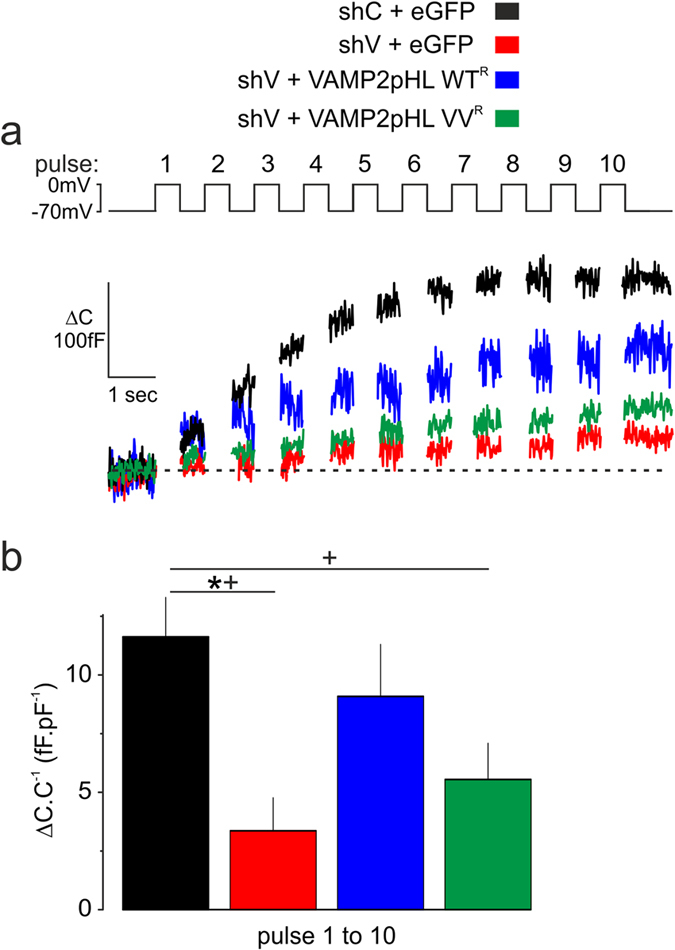
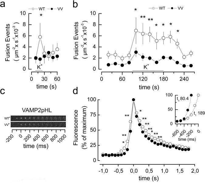
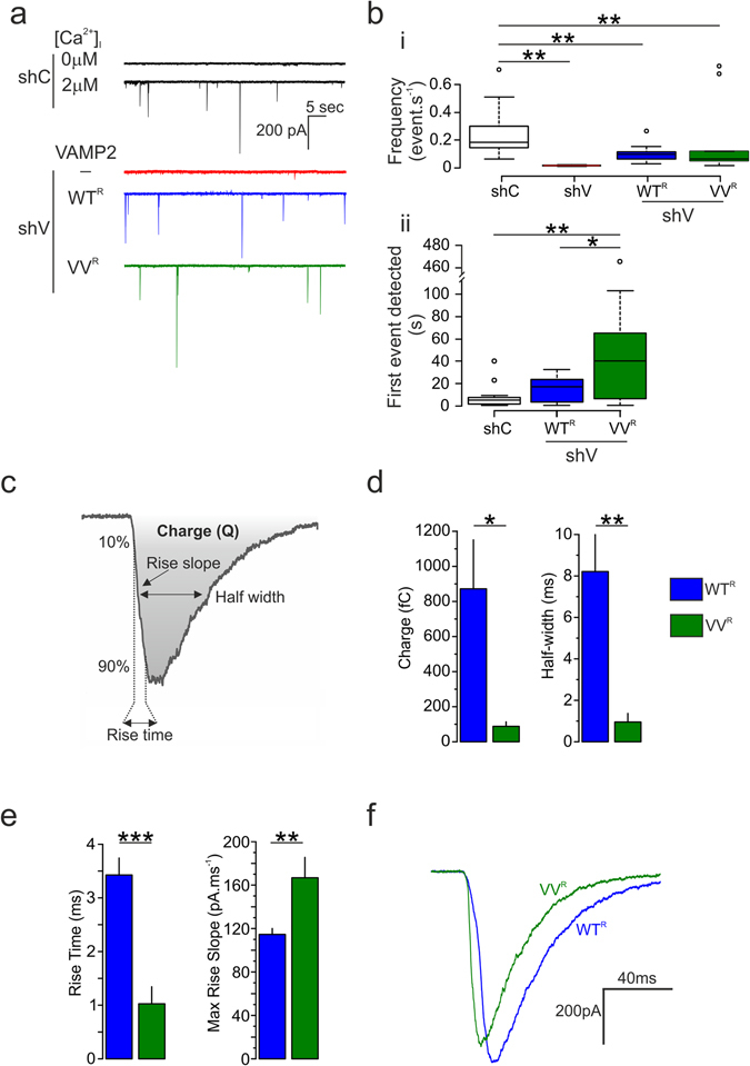
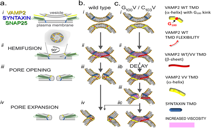
Exocytosis depends on cytosolic domains of SNARE proteins but the function of the transmembrane domains (TMDs) in membrane fusion remains controversial. The TMD of the SNARE protein synaptobrevin2/VAMP2 contains two highly conserved small amino acids, G100 and C103, in its central portion. Substituting G100 and/or C103 with the β-branched amino acid valine impairs the structural flexibility of the TMD in terms of α-helix/β-sheet transitions in model membranes (measured by infrared reflection-absorption or evanescent wave spectroscopy) during increase in protein/lipid ratios, a parameter expected to be altered by recruitment of SNAREs at fusion sites. This structural change is accompanied by reduced membrane fluidity (measured by infrared ellipsometry). The G100V/C103V mutation nearly abolishes depolarization-evoked exocytosis (measured by membrane capacitance) and hormone secretion (measured biochemically). Single-vesicle optical (by TIRF microscopy) and biophysical measurements of ATP release indicate that G100V/C103V retards initial fusion-pore opening, hinders its expansion and leads to premature closure in most instances. We conclude that the TMD of VAMP2 plays a critical role in membrane fusion and that the structural mobility provided by the central small amino acids is crucial for exocytosis by influencing the molecular re-arrangements of the lipid membrane that are necessary for fusion pore opening and expansion.


Conflict of interest statement

The authors declare that they have no competing interests.

Figures

Figure 1
Figure 1
Conserved small residues in the N-terminal portion of the transmembrane domain of VAMP2 have a role in exocytosis. (a) Phylogenetic comparison of the VAMP2 transmembrane domain. The highly conserved small amino acids at the position 100 and 103 are highlighted in red. In the consensus sequence, residues that localize on the same aspect of the helical structure as G100 and C103 are presented in yellow. (b) Helical wheel presentation of the N-terminal half of the transmembrane domain highlighting the clustering of small (red) to medium-sized residues on a single face. (c) Sequences of wild-type and mutant TMDs; point mutations are highlighted in red. Mutations of small residues in the transmembrane domain (TMD) inhibit secretion in PC12 cells. Effects on hormone secretion were determined for mutants of the VAMP2 TMD after knock-down of endogenous VAMP2. (d) Transient expression of shRNA against VAMP2 (shV) but not control shRNA (shC) reduces expression of VAMP2 in PC12 cells. Tubulin immunoreactivity is shown for comparison. Full blots are given in Supplemental Fig. 8. (e) Quantification of silencing efficiency of shC and shV on endogenous VAMP2 as indicated. Data have been normalised to control conditions (no transfection). N = 3, **p < 0.01. (f) Re-expression of shRNA-resistant VAMP2. Cells were co-transfected with a plasmid bearing shC or shV, a bicistronic plasmid expressing either eGFP alone (pPRIG empty) or both eGFP and VAMP2. Wild-type VAMP2 was either sensitive (WTS) or resistant (WTR) to shV. All mutants were resistant to shV (VAMP2R). Full blots are given in Supplemental Fig. 8. (g) Quantification of re-expression of exogenous VAMP2Rs normalised to control (WTR). N = 3. (h) Reconstitution of depolarization-induced secretion. PC12 cells were transiently co-transfected with a plasmid expressing the human growth hormone (hGH), shC or shV, and the indicated VAMP2R (WTR or mutant in the TMD). Basal and stimulated hGH secretion are given. Mean values ± S.E.M., N as indicated. *p < 0.05, **p < 0.01 (ANOVA and Bonferroni). (i) Same data as g presented as percentage of reconstitution of stimulated secretion. Statistics as in h.
Figure 2
Figure 2
The VV mutation reduces structural flexibility of the VAMP2 TMD. (a) Sequences and space-filling models of the transmembrane domain of the WT and VV mutant VAMP2 as an α-helix. Yellow designates sulphur, green carbon, white hydrogen and red oxygen. Arrows designate positions of G100 and C103, arrowheads point to the volume changes in the mutated sites. (b) ATR–IR spectra of the synthetic peptide VAMP295-116 (WT or VV mutant) in a lipid multi-bilayer (DOPC) were obtained at a peptide/lipid ratio of 1/20 at room temperature. After 1 h peptides were diluted with DOPC to a peptide/lipid ratio of 1/250 and structural changes measured for 2 h at room temperature. The ratios of α-helices vs β-sheets are given for wild-type (○) or the VV mutant (●). The curves were obtained fitting exponential growth. The time constants (τ) are given next to the curves.
Figure 3
Figure 3
The VV mutations in VAMP2 TMD reduce structural dynamics. Structural features of VAMP2 WT or VAMP2 VV full-length proteins were measured by infrared spectroscopy (PMIRRAS) in a Langmuir through at different lateral pressure. Note that increases in pressure increase local protein concentrations and peptide/lipid ratios. All experiments were performed at room temperature. Full-length proteins were embedded in DMPC monolayer at the protein/lipid ratio of 1/50 (Comparable results were obtained at peptide/lipid ratios of 1/20, data not shown). α-helical conformations and β-sheets conformations were detected at 1653 cm−1 and 1630 cm−1, respectively. (a) Structural behaviour of either VAMP2 WT or VV-TMDs during compression of the DMPC monolayer (ai and aii, respectively). (a i) Starting from a low lateral pressure (5 mN.m−1, black curve), a shoulder at 1653 cm−1 (corresponding to α helices) was detected. During compression to 44 mN.m−1, this shoulder becomes less prominent (red curve, arrowhead). (a ii) Identical procedure performed with VV from 4 mN.m−1 (black curve) to 55 mN.m−1 (red curve). Note the persistence of the shoulder (arrowhead) at high pressure. (b) Structural behaviour of either VAMP2 WT or VV-TMDs during decompression of the DMPC monolayer (bi and bii, respectively). (b i) To facilitate comparison, the high pressure curve from A has been superimposed (red traces). Decompression of the monolayer from 44 to 4 mN.m−1 restores the α helix shoulder (bi, blue curve, arrowhead), whereas decompression from 55 mN.m−1 to 4 mN.m−1 on membrane containing VV does not change the absorbance curve (b ii, blue curve and arrowhead).
Figure 4
Figure 4
The VV mutation in VAMP2 TMD modifies the fluidity of the membrane. Viscosity of model membranes were imaged by ellipsometry in a Langmuir through using DMPC membranes and either VAMP2 WT or VAMP2 VV full-length recombinant protein. (a) Representative images from DMPC model membranes mixed with the indicated mutant (1/50 nominal peptide/lipid ratio) obtained by ellipsometry in a Langmuir trough. Images were taken at initial low lateral pressure (left panel), at maximal lateral pressure (middle panel) and after relaxation (to low pressure, right panel). Measured lateral pressures are given at the bottom left corner of each image (mN/m). For VAMP2 WT, during the increase of the lateral pressure, the DMPC membrane evolves from homogenous monolayer to a monolayer bearing distinctive domains of different thickness (clear and dark zones). At the maximal pressure (36 mN/m), the patterns (round shapes, no sharp angles) indicate regions of great fluidity. By decompressing the system, the membrane returns fully to its original homogeneity. By contrast, an increase of the lateral pressure on membrane containing VV leads to the formation of ‘jagged’ patches with many sharp angles, a mark of membrane rigidity (28 mN/m). These changes persist upon decompression. (b) Quantification of fractional dimension of images obtained in ellipsometry by using the box counting dimension (mean DB, a logarithmic factor which varies between 1 and 2). Mean ± S.E.M., n = 6; **2p < 0.01 (t-test).
Figure 5
Figure 5
Effects of mutant VAMP2 TMDs on exocytosis measured by membrane capacitance in INS-1 832/13 clonal β-cells. Reconstitution of exocytosis by VAMP2 WT or VAMP2 VV and kinetics of vesicle pools were measured in INS-1 832/13 clonal β-cells expressing VAMP2pHL WTR or VAMP2pHL VVR after knock-down of endogenous VAMP2. (a) Representative traces of cumulative increase in membrane capacitance (ΔC in fF) elicited by 10 depolarizations (top panel) from −70 mV to 0 mV applied at 1 Hz. Cells were co-transfected with either shC + eGFP (n = 5, black trace), shV + eGFP (n = 6, red trace), shV + VAMP2pHL WTR (n = 6, blue trace) or shV + VAMP2pHL VVR (n = 10, green trace). (b) Quantification of the cumulative increase of capacitance normalized to cell capacitance (ΔC.C−1) at the end of the train. Although presenting a clear trend, the difference in the cumulative exocytosis measured in cells expressing either VAMP2pHL WTR or VAMP2pHL VVR did not reach statistical significance (ANOVA and Tukey, p = 0.093, *p < 0.05). However, differences became significantly evident when comparing the kinetics of exocytosis for WTR or VVR to those of the reference control shC (ANOVA and Dunnett, +, p < 0.05).
Figure 6
Figure 6
The VV mutation in the TMD of VAMP2 alters exocytosis and fusion pore kinetics. Exocytosis and release kinetics were determined by near-field TIRF microscopy in PC12 and in INS-1 832/13 clonal β-cells expressing VAMP2 WT or the VAMP2 VV mutant after knock-down of endogenous VAMP2. (a) PC12 cells were transiently co-transfected with plasmids encoding VAMP2-pHluorin (WTR or VVR) and shRNA directed against VAMP2. Fusion events were recorded by TIRF microscopy and sum of fusion events per 10 s is given for cells stimulated by 90 mM [K+]o (WT, n = 6 cells, 237 events; VV n = 4 cells, 139 events total) *2p < 0.05 (Student’s t-test). (b) Same as in (a) but measurements performed on INS-1 832/13. Sum of fusion event per 20 s in transfected cells were stimulated by 35 mM [K+]o for 30 s (WT n = 9 cells, 1114 events total; VV n = 9 cells, 476 events total. *2p < 0.05; **2p < 0.01 (Student’s t-test). (c) Representative films of fusion events in INS-1 832/13 cells. The events are temporally synchronized to the frame of their maximum fluorescence. (d) Mean changes in normalized fluorescence immediately before and after exocytosis in INS-1 832/13 cells expressing either VAMP2pHL WTR (open circles) or VVR (closed circles). For display the fluorescent mean events were synchronized to their maximum fluorescence. Note narrower peak for VV as compared to WT. *2p < 0.05; **2p < 0.01 VAMP2 WT vs. VV (Student’s t-test). INSERT: Events synchronized for first event with a mean >10% of maximal fluorescence and curves fitted for first order exponential growth. Note shorter time constant τ for VV (closed circles) as compared to WT (open circles). The time constants τup for the fluorescence increases averaged 189 ± 28 ms for WTR and 80 ± 7 ms for VVR (p < 0.05; F-test). Similarly, the decay of fluorescence following the peak was slower for WTR (time constant τdown: 558 ± 57 ms) than for VVRdown: 315 ± 17 ms) (p < 0.05; F-test).
Figure 7
Figure 7
VAMP2 G100/C103 influence fusion pore opening. Measurements of quantal release of ATP via P2X2 receptor mediated currents from INS-1 832/13 cells expressing VAMP2 WT (G100/C103) or VV (G100V/C103V) after knock-down of endogenous VAMP2. (a) Currents triggered by ATP release. Top traces show INS-1 832/13 cells transfected with shC (black trace) and infused with media containing 0 or 2 μM free calcium. The 3 bottom traces are from cells co-transfected with plasmid P2X2-YFP, shC or shV, and VAMP2-pHL (WTR or VVR), respectively (black, shC; red, shV + empty vector; blue, shV + VAMP2pHL WTR; green; shV + VAMP2pHL VVR). (b i) Frequency of events per second (ANOVA and Tukey shC vs. shV p = 0.004, vs WTR p = 0.004, vs. VVR p = 0.001; shC, 12 cells, n = 802 events total; shV, 6 cells, n = 30 events total; WTR 12 cells, n = 388 events; VVR, 8 cells, n = 695 events; °, outliers). (b) Response delay defined as time required for the first event to be detected (ANOVA and Tukey, shC = 4 ± 0.86 s, WTR = 14, 43 ± 3.21 s, VVR = 36.90 ± 11.48 s. (c) Schematic illustration of the charge, the 10% to 90% rise time, the rise slope, and the half width of the current generated by P2X2R. These parameters are measured for the fusion pore kinetics study. (d,e) Mean values of the medians for the charge, the half width (d) and rise time and its slope (e), respectively (*p < 0.05, **p < 0.01, t test). (f) Superimposition of currents of similar amplitude elicited from cells expressing VAMP2pHL WTR (blue) and VVR (green).
Figure 8
Figure 8
Proposed model of conformation of VAMP2 transmembrane domain during exocytosis. The different steps of exocytosis are viewed from general scale with the presence of the SNARE cytosolic domain (a) and more focused on the TMD structure from VAMP2 WT (b) and VAMP2 VV (G100V/C103V) (c). Before vesicle docking, VAMP2 is uniformly distributed over the vesicle, corresponding to a low local concentration of VAMP2, and the TMDs are mainly in α-helical conformation (a i,b i, c i). Following docking, VAMP2 proteins concentrate at the fusion site (a ii). The increased local TMD concentration alters the peptide/lipid ratio and induces the switch of its conformation from an α-helical to a β-sheet conformation. The structural change is accompanied by a tilt of angle of the transmembrane domain from 30° to 54°, promoting the lipid reorganisation and an increase of membrane viscosity (b ii,c ii). Once fusion has occurred and following lipid rearrangement, the SNAREs are localised on the same membrane (a iii). The decrease of the local concentration of VAMP2 and membrane tension results in the return to an α-helical conformation in the case of VAMP2 WT. This is accompanied by an increase of fluidity of the membrane that promotes the opening of the fusion pore for VAMP2 WT (b iii) and its subsequent expansion (a iv and b iv). The VV mutant has reduced capacity for reversible structural changes and is either lacks flexibility around G100 or is locked in the β-sheet conformation. This causes a delay in the fusion pore opening in the case of VAMP2 VV (c iib, c iii). The persisting increase in membrane viscosity does not permit further expansion of the pore and induces its premature closure in most cases (c iic). In some cases full fusion may occur as documented by recording of membrane capacitance.

Similar articles

Cited by

References

    1. Rothman JE. The principle of membrane fusion in the cell (Nobel lecture) Angew. Chem. Int. Ed. Engl. 2014;53:12676–12694. doi: 10.1002/anie.201402380. - DOI - PubMed
    1. Jahn R, Fasshauer D. Molecular machines governing exocytosis of synaptic vesicles. Nature. 2012;490:201–207. doi: 10.1038/nature11320. - DOI - PMC - PubMed
    1. Chernomordik LV, Kozlov MM. Mechanics of membrane fusion. Nat. Struct. Mol. Biol. 2008;15:675–683. doi: 10.1038/nsmb.1455. - DOI - PMC - PubMed
    1. Hernandez JM, Kreutzberger AJ, Kiessling V, Tamm LK, Jahn R. Variable cooperativity in SNARE-mediated membrane fusion. Proc. Nat’l Acad. Sci. USA. 2014;111:12037–12042. doi: 10.1073/pnas.1407435111. - DOI - PMC - PubMed
    1. Kozlov MM, Chernomordik LV. Membrane tension and membrane fusion. Curr. Opin. Struct. Biol. 2015;33:61–67. doi: 10.1016/j.sbi.2015.07.010. - DOI - PMC - PubMed

Publication types

Substances

LinkOut - more resources