. 2017 Jun 6;7(1):2811.
doi: 10.1038/s41598-017-02557-8.

Irisin prevents and restores bone loss and muscle atrophy in hind-limb suspended mice

Affiliations

Irisin prevents and restores bone loss and muscle atrophy in hind-limb suspended mice

Graziana Colaianni et al. Sci Rep. .

Abstract

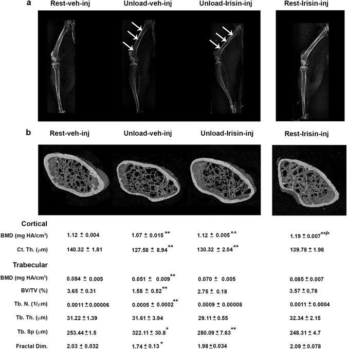
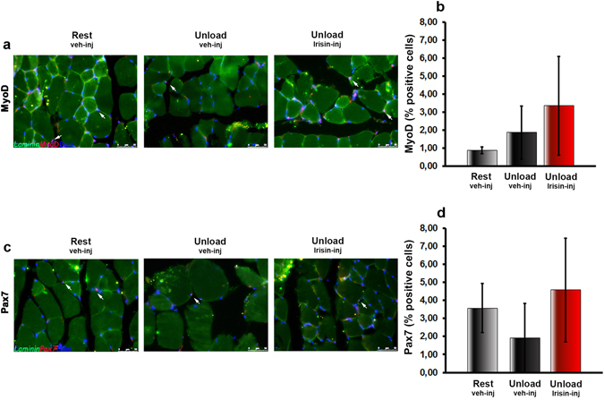
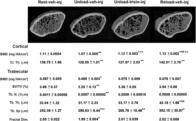
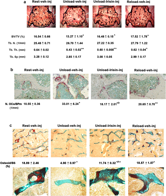
We previously showed that Irisin, a myokine released from skeletal muscle after physical exercise, plays a central role in the control of bone mass. Here we report that treatment with recombinant Irisin prevented bone loss in hind-limb suspended mice when administered during suspension (preventive protocol) and induced recovery of bone mass when mice were injected after bone loss due to a suspension period of 4 weeks (curative protocol). MicroCT analysis of femurs showed that r-Irisin preserved both cortical and trabecular bone mineral density, and prevented a dramatic decrease of the trabecular bone volume fraction. Moreover, r-Irisin protected against muscle mass decline in the hind-limb suspended mice, and maintained the fiber cross-sectional area. Notably, the decrease of myosin type II expression in unloaded mice was completely prevented by r-Irisin administration. Our data reveal for the first time that Irisin retrieves disuse-induced bone loss and muscle atrophy. These findings may lead to development of an Irisin-based therapy for elderly immobile osteoporotic and physically disable patients, and might represent a countermeasure for astronauts subjected to microgravity-induced bone and muscle losses.


Conflict of interest statement

M. Grano, S. Cinti, G. Colaianni, C. Cuscito, S. Colucci, G. Mori, G. Brunetti are name inventors of the Italian patent (MI2015A000558) and the European patent (16165324.1-1453) titled “Irisin for care and prevention of osteoporosis” related to the work described.

Figures

Figure 1
Figure 1
Treatment with r-Irisin prevents bone loss in femurs from hindlimb-suspended mice. (a) Contact radiographs of selected long bones from normal loading mice (Rest veh-inj) and unloaded mice treated with vehicle or recombinant Irisin (r-Irisin, 100 µg kg−1 per week for 28 days, mice were sacrificed 24 hours after last dose). Arrows indicate difference in radiodensity between femur of unloaded mice treated with vehicle and femur of unloaded mice treated with recombinant Irisin. (b) Representative micro-CT-generated section images and calculated cortical and trabecular parameters of femurs harvested from Rest mice vehicle- or Irisin-injected and Unload mice vehicle- or r-Irisin-injected. Cortical bone parameters included bone mineral density (BMD) and cortical thickness (Ct.Th). Trabecular bone parameters included bone mineral density (BMD), bone volume/total volume (BV/TV), trabecular number (Tb.N), trabecular thickness (Tb.Th), trabecular separation (Tb Sp) and Fractal Dimension. Data are presented as mean ± SEM. n = 7–8 mice per group. All data were normally distributed according to the Shapiro-Wilk normality test and analyzed by one-way ANOVA and Bonferroni’s post hoc analysis. Cohen’s d values were measured for non-significant differences of results and can be found as Supplementary Table S1. *p ≤ 0.05, **p ≤ 0.01 versus Rest vehicle-injected mice. ^ p ≤ 0.05, ^^ p ≤ 0.01 versus Unload vehicle-injected mice.
Figure 2
Figure 2
Treatment with r-Irisin inhibits sclerostin increase and Opg decrease caused by unloading in vivo. (a) Western immunoblotting and densitometric quantitation of sclerostin expression versus control loading (β-actin) in long bones (depleted of bone marrow) harvested from Rest vehicle-injected and Unload vehicle- or r-Irisin-injected mice (r-Irisin, 100 µg kg−1 per week for 28 days, mice sacrificed 24 hours after last dose). (b) Opg and (c) Rank-l mRNA expression (qPCR) in long bones (depleted of bone marrow) harvested from Rest vehicle-injected and Unload vehicle- or r-Irisin-injected mice. (d) Ex vivo Cfu-f formation (%), (e) Alp and (f) Collagen I mRNA expression (qPCR) in ex vivo cultures obtained from bone marrow harvested from Rest vehicle-injected and Unload vehicle- or r-Irisin-injected mice. (g) TRAP-positive osteoclast formation (%), (h) Trap and (I) Cathepsin K mRNA expression (qPCR) in ex vivo cultures obtained from bone marrow harvested from Rest veh-injected and Unload vehicle- or r-Irisin-injected mice. Data are presented as mean ± SEM. n = 3 mice per group. All data were normally distributed according to the Shapiro-Wilk normality test and analyzed by one-way ANOVA and Bonferroni’s post hoc analysis. *p ≤ 0.05, **p ≤ 0.01 versus Rest vehicle-injected mice. ^ p ≤ 0.05 versus Unload vehicle-injected mice.
Figure 3
Figure 3
Treatment with r-Irisin prevents muscle wasting in hindlimb-suspended mice. (a) Vastus lateralis weight normalized to total body weight from normal loading mice (Rest veh-inj) and unloaded mice treated with vehicle or recombinant Irisin (r-Irisin, 100 µg kg−1 per week for 28 days, mice sacrificed 24 hours after last dose). (b) Photomicrographs of hematoxylin and eosin stained sections of vastus lateralis from Rest vehicle-injected and Unload vehicle- or Irisin-injected mice (magnification: 20x). (c) Quantitative assessments of Cross-Sectional Area (CSA) and (d) CSA area distribution of fibers from vastus lateralis harvested from normal loading mice (Rest vehicle-inj) and unloaded mice treated with vehicle or r-Irisin. Data are presented as mean ± SEM. n = 6–7 mice per group. All data were normally distributed according to the Shapiro-Wilk normality test and analyzed by one-way ANOVA and Bonferroni’s post hoc analysis. *p ≤ 0.05, ***p ≤ 0.001 versus Rest vehicle-injected mice. ^ p ≤ 0.05 versus Unload vehicle-injected mice.
Figure 4
Figure 4
Treatment with r-Irisin inhibits myosin heavy chain type II decrease caused by unloading in vivo. (a) Representative images of immunohistochemistry staining of VDAC protein in vastus lateralis from normal loading mice (Rest vehicle-inj) and unloaded mice treated with vehicle or recombinant Irisin (r-Irisin, 100 µg kg−1 per week for 28 days, mice sacrificed 24 hours after last dose) (magnification: 40x). (b) Quantitative assessment of percentage of VDAC staining. (c) Densitometric quantitation of cytomchrome c oxidase subunit I (COX IV) expression versus control loading (α-tubulin) in vastus lateralis isolated from Rest vehicle-injected and Unload vehicle- or r-Irisin-injected mice. (d) Nrf-1 and (e) Tfam mRNA expression (qPCR) in vastus lateralis harvested from Rest vehicle-injected and Unload vehicle- or Irisin-injected mice. (f) Electron microscope images. (g) Fluorescent micrographs of vastus lateralis sections of Rest vehicle-injected and Unload vehicle- or r-Irisin-injected mice immunolabeled for FNDC5 (green) and ATP synthase (red) (magnification: 20x). (h) Quantitative assessment of percentage of co-localization of FNDC5 and ATP synthase positive fibers (i) Densitometric quantitation of myosin heavy chain type II (MyHC II) expression versus control loading (α-tubulin) in vastus lateralis isolated from Rest vehicle-injected and Unload vehicle- or r-Irisin-injected mice. (j) MyHC IIx, (k) MyHC IIβ and (l) MyHC I mRNA expression (qPCR) in vastus lateralis harvested from Rest vehicle-injected and Unload vehicle- or Irisin-injected mice. Data are presented as mean ± SEM. n = 3–4 mice per group. All data were normally distributed according to the Shapiro-Wilk normality test. Results from Fig. 4 were analyzed by one-way ANOVA and Bonferroni’s post hoc analysis. *p ≤ 0.05, **p ≤ 0.01, ***p ≤ 0.001 versus Rest vehicle-injected mice. ^ p ≤ 0.05, ^^^ p ≤ 0.001 versus Unload vehicle-injected mice.
Figure 5
Figure 5
Treatment with r-Irisin does not affect satellite cells in hindlimb-suspended mice. Fluorescent micrographs of vastus lateralis sections from Rest vehicle-injected and Unload vehicle- or Irisin-injected mice, immunostained (red) for MyoD (a) and Pax7 (c) and counterstained for Laminin (green) and DAPI (blue). The percentage of MyoD positive (b) and Pax7 positive (d) cells are quantified. Data are presented as mean ± SEM. n = 3 mice per group. All data were normally distributed according to the Shapiro-Wilk normality test and analyzed by one-way ANOVA and Bonferroni’s post hoc analysis.
Figure 6
Figure 6
Treatment with r-Irisin recovers bone loss in femurs from hindlimb-suspended mice. Representative microCT-generated section images and calculated cortical and trabecular parameters of femurs obtained from normal loading mice (Rest vehicle-inj), unloaded mice treated with vehicle or recombinant Irisin (first r-Irisin injection after 4 weeks of hindlimb suspension, at the dose of 100 µg kg−1 per week for 28 days) and reloaded mice (Reload veh-inj). Cortical bone parameters included bone mineral density (BMD) and cortical thickness (Ct.Th). Trabecular bone parameters included bone mineral density (BMD), bone volume/total volume (BV/TV), trabecular number (Tb.N), trabecular thickness (Tb.Th), trabecular separation (Tb Sp) and Fractal Dimension. Data are presented as mean ± SEM. n = 6–7 mice per group. All data were normally distributed according to the Shapiro-Wilk normality test and analyzed by one-way ANOVA and Bonferroni’s post hoc analysis. Cohen’s d values were measured for non-significant differences of results and can be found as Supplementary Table S1. *p ≤ 0.05, **p ≤ 0.01, ***p ≤ 0.001 versus Rest vehicle-injected mice. ^ p ≤ 0.05, ^^ p ≤ 0.01, ^^^ p ≤ 0.001 versus Unload vehicle-injected mice.
Figure 7
Figure 7
Treatment with r-Irisin recovers bone loss in vertebrae from hindlimb-suspended mice. (a) Von Kossa-stained vertebral sections and trabecular bone parameters from normal loading mice (Rest vehicle-inj), unloaded mice treated with vehicle or recombinant Irisin (first r-Irisin injection after 4 weeks of hindlimb suspension, at the dose of 100 µg kg−1 per week for 28 days) and reloaded mice (Reload vehicle-inj) (magnification: 2.5x). (b) Representative images of tartrate-resistant acid phosphatase-stained osteoclasts in vertebral sections, together with osteoclast counts per bone perimeter (BPm). (magnification: 40x). (c) Representative images of Goldner’s Masson Trichrome-stained vertebral sections (magnification: 10x) and photomicrograph of details taken at 40x, together with measurement of percentage of osteoid per bone surface (BS). n = 4–5 mice per group. Data are presented as mean ± SEM. All data were normally distributed according to the Shapiro-Wilk normality test and analyzed by one-way ANOVA and Bonferroni’s post hoc analysis. *p ≤ 0.05, **p ≤ 0.01 versus Rest vehicle-injected mice. ^ p ≤ 0.05, ^^ p ≤ 0.01 versus Unload vehicle-injected mice.

References

    1. Zehnder Y, et al. Long-term changes in bone metabolism, bone mineral density, quantitative ultrasound parameters, and fracture incidence after spinal cord injury: a cross-sectional observational study in 100 paraplegic men. Osteoporos Int. 2004;15(3):180–9. doi: 10.1007/s00198-003-1529-6. - DOI - PubMed
    1. Jørgensen L, Jacobsen BK, Wilsgaard T, Magnus JH. Walking after stroke: does it matter? Changes in bone mineral density within the first 12 months after stroke. A longitudinal study. Osteoporosis Int. 2000;11:381–7. doi: 10.1007/s001980070103. - DOI - PubMed
    1. Oppl B, et al. Low bone mineral density and fragility fractures in permanent vegetative state patients. J Bone Miner Res. 2014;29(5):1096–100. doi: 10.1002/jbmr.2122. - DOI - PubMed
    1. Takamoto S, et al. Spontaneous fractures of long bones associated with joint contractures in bedridden elderly inpatients: clinical features and outcome. J Am Geriatr Soc. 2005;53(8):1439–41. doi: 10.1111/j.1532-5415.2005.53433_3.x. - DOI - PubMed
    1. LeBlanc A, et al. Bone mineral and lean tissue loss after long duration space flight. J Musculoskelet Neuronal Interact. 2000;1(2):157–60. - PubMed

Publication types

MeSH terms

LinkOut - more resources