Review
. 2017 May;473(2201):20160822.
doi: 10.1098/rspa.2016.0822. Epub 2017 May 31.

Quantum effects in biology: golden rule in enzymes, olfaction, photosynthesis and magnetodetection

Affiliations
Review

Quantum effects in biology: golden rule in enzymes, olfaction, photosynthesis and magnetodetection

Jennifer C Brookes. Proc Math Phys Eng Sci. 2017 May.

Abstract

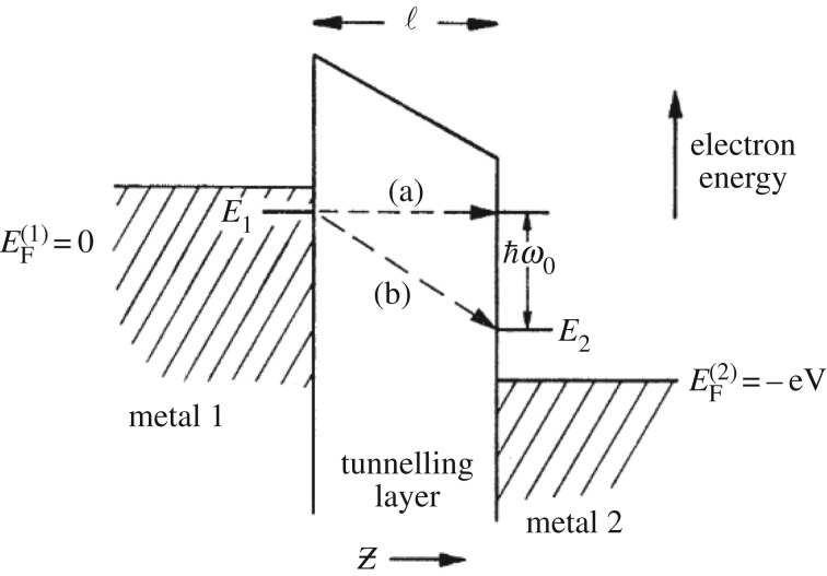
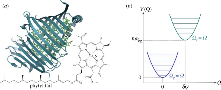
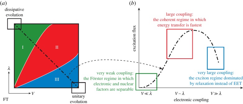
Despite certain quantum concepts, such as superposition states, entanglement, 'spooky action at a distance' and tunnelling through insulating walls, being somewhat counterintuitive, they are no doubt extremely useful constructs in theoretical and experimental physics. More uncertain, however, is whether or not these concepts are fundamental to biology and living processes. Of course, at the fundamental level all things are quantum, because all things are built from the quantized states and rules that govern atoms. But when does the quantum mechanical toolkit become the best tool for the job? This review looks at four areas of 'quantum effects in biology'. These are biosystems that are very diverse in detail but possess some commonality. They are all (i) effects in biology: rates of a signal (or information) that can be calculated from a form of the 'golden rule' and (ii) they are all protein-pigment (or ligand) complex systems. It is shown, beginning with the rate equation, that all these systems may contain some degree of quantumeffect, and where experimental evidence is available, it is explored to determine how the quantum analysis aids in understanding of the process.

Keywords: enzymes; magnetodetection; olfaction; photosynthesis; quantum.


Conflict of interest statement

I declare I have no competing interests.

Figures

Figure 1.
Figure 1.
(a) A configuration coordinate diagram showing the activation energy barrier E, separating the reactant states (donor D) from the product states (acceptor A) which are shown as diabatic Born–Oppenheimer surfaces, with κ=1, as in equation (1.2). Shown also, ΔE the energy difference at minimum between the D and A, at XD and XA, respectively, λ the reorganization energy and the reaction coordinate Q [2,1]. Xc indicates a crossing point for the transitioning particle at the asterisk. (b) a classical tunnelling (or insulating) barrier that separates the donor and acceptor positions, barrier height is E, barrier width is distance r.
Figure 2.
Figure 2.
DeVault & Chance's pictorial description of the tunnelling region between cytochrome and bacteriochlorophyll (BChl) from [9] with permission from Elsevier. Energetic levels occupied by valence electrons are shown. The BChl is excited by light v which leaves a hole quickly filled by an electron tunnelling from the cytochrome. DeVault & Chance also indicate the possibility of an ‘activated’ path versus the tunnelling path, where the activated path may be enabled, for example by a vibration in the cytochrome, bringing together the energetic states to the crossing point (asterisk, see figure 1) and/or the tunnelling path also may be ‘stabilized by a phonon emission’ [9].
Figure 3.
Figure 3.
Showing the internal hydrogen transfer in malonaldehyde from Hay & Scrutton [14]. (a,b) The transition towards product from reactant, via the transition state (TS). (c) Donor (D) and acceptor (A) moving along a reaction coordinate, Q, the dashed line shows where tunnelling is most probable at the TS. The TRC denotes ‘tunnelling ready coordinate’. (d) The potential energy surface along a reaction coordinate for hydrogen and deuterium transfer at the same D–A distances. The idea of a ‘promoting mode’: to enhance the hydrogen transition is depicted, if motion in c d(Q) is conducive to promotion, then the tunnelling distance dtun is reduced and in combination with a lowering of the activation enthalpy a rate enhancement results.
Figure 4.
Figure 4.
Depiction of vibrationally assisted models of hydrogen transfer from [14]. E denotes energy on the potential energy surface, q is the environmental reaction coordinate, r is the hydrogen coordinate where transfer is indicated by the red sphere, occurring at crossing point q*. qR denotes hydrogen in the initial/D state, and qP the hydrogen on the final/ A state. ‘M’ denotes this Marcus-like depiction, see equation (2.2), of the Born–Oppenheimer states on the environmental reaction coordinate. In a promoting mode, a vibrational motion must be coupled to the itinerant charge. (Online version in colour.)
Figure 5.
Figure 5.
Lambe & Jacklevic demonstrate inelastic electron tunnelling through a metal junction, from [32]. There are two possible ways for the electron to cross the tunnelling layer (insulating barrier) via (a) an elastic or (b) an inelastic transition. The inelastic transition occurs when there is a molecule bridging the tunnelling layer with a mode of vibration, ωo, that the tunnelling electrons excites, and so loses energy too.
Figure 6.
Figure 6.
IR spectra for ‘4-d30’ (red line) and undeuterated muscone ‘4’ (black line) indicating differing vibrational modes (particularly in the 1380–1550 cm−1 region) from [45] copyright Proceedings of National Academy of Sciences, USA. Also shown are dose-response curves of OR5AN1 plots for (1) cyclopentadecanone and (4) muscone and the scatter plots with 96% confidence interval log EC50 values, showing there is no differentiation between isotopes that is statistically significant [45].
Figure 7.
Figure 7.
(a) A PPC from pdb code 3BSD showing the seven pigments in the Fenna–Matthews–Olson (FMO) complex in Chlorobaculum tepidum (formerly Chlorobium tepidum) from [55]. The structure of the FMO is shown in blue ribbon and the chromophores: bacteriochlorophyll a (BChl a) is in green. The two-dimensional molecular structure of the seven BChl a is also shown next to the protein [55]. (b) Born–Oppenheimer surfaces to show the shift from ground to excited state on a BChl a pigment in FMO [56].
Figure 8.
Figure 8.
Figure to show the incoherent Förster (very weak coupling), intermediate coherent (large coupling) and Redfield (very large coupling) regime for EET in photosynthesis, from [58]. The electronic coupling refers to an electrostatic (Coloumb) dipole–dipole interaction between pigment BChl sites.
Figure 9.
Figure 9.
Two-dimensional electronic spectroscopy (2DES), in (a) showing the time evolution of excitation between 2 pigments, ‘cross peaks’ amplitudes oscillate with v2v1, Fourier transform gives the oscillation frequency. (b) shows the two-dimensional spectra at T=55 fs (real part of total signal) for PC645, a marine cryptophyte algae that contains 8 billion (chromophore) pigments, coloured bars are peak absorptions for the chromophores. The green cross indicates a cross peak with coherent oscillation as a function of waiting time T (at 570 nm, 600 nm). (c) plots the oscillation's amplitude with waiting time (d) plots the rephasing (red) and non-rephasing (purple) amplitudes of this cross peak and (e) is a Fourier transform of the traces in (d) showing that peaks occur at 26 THz in both signals but 21 THz only in the rephasing spectrum, from [58].
Figure 10.
Figure 10.
The first evidence of electronic coherence between single states in FMO, a non-rephasing signal appears on the main diagonal in (a) and evolved in time, see (b). Plotting the amplitude of the lowest exciton peak beating with time, upon Fourier transform (d) reproduces frequencies that agree well with the predicted beat times and intensities, from [62].
Figure 11.
Figure 11.
Quantum beats at room temperature in FMO from [64], copyright Proceedings of National Academy of Sciences, USA. Data shown are at waiting time T=400 fs and (a) is 77 K (b) 125 K (c) 150 K and (d) 277 K. At higher temperature, peaks are broader (dephasing between g and e states). Theoretical analysis [65], extracts the quantum beat at the cross peak (white circle) and plotted in (e) as a function of waiting time T to observe the amplitude oscillating. The beat occurs around 200 fs even at room temperature.
Figure 12.
Figure 12.
Experiments show birds exposed to monochromatic light exhibit differing orientation behaviour—orientation is better in ‘blue’ light, from [81].
Figure 13.
Figure 13.
RPM mechanism adapted from [89]. The left shows the photo-initiated radical pair AB (FH and W here), the spin Hamiltonian H^ interconverts the pair from singlet to triplet states, with differing resulting singlet or triplet products as a consequence of the entanglement in the middle: structures in the box show the tryptophan radical (W) and the flavin radical (FH) at the proposed orientation in vivo, (the possible AB pair). On the left are the two-dimensional structures for FH and W. The orientation of this possible RP (from pdb code 1DNP) was used to calculate anisotropic magnetic field effects in [89].
Figure 14.
Figure 14.
The orientation dependence of the singlet recombination probability ΦS of the radical pair [FH•++W•−] as in figure 13, from [89]. Results are for a weak field of B=50 μT and kS=2×10⁵ s−1.
Figure 15.
Figure 15.
Reaction mechanism (b) and two-dimensional structures of the carotenoid (C)–porphyrin (P)–fullerene (F) system (a) that demonstrates the ‘chemical compass’ principle, from [91]. Interconversion between singlet and triplet pair is shown [C•+PF•−] as moderated by weak magnetic fields. The rates at which they recombine to C–P–F is spin-selective accounting for rates kS and kT. C•+ shows the anisotropic hyperfine couplings calculated on C.

References

    1. De Vault D. 1984. Quantum-mechanical tunneling in biological systems, vol. 2 Cambridge, UK: Cambridge University Press.
    1. Klinman JP, Kohen A. 2013. Hydrogen tunneling links protein dynamics to enzyme catalysis. Annu. Rev. Biochem. 82, 471–496. (doi:10.1146/annurev-biochem-051710-133623) - DOI - PMC - PubMed
    1. Eyring H. 1935. The activated complex and the absolute rate of chemical reactions. Chem. Rev. 17, 65–77. (doi:10.1021/cr60056a006) - DOI
    1. Gray HB, Winkler JR. 2005. Long-range electron transfer. Proc. Natl Acad. Sci. USA 102, 3534–3539. (doi:10.1073/pnas.0408029102) - DOI - PMC - PubMed
    1. Oppenheimer JR. 1928. Three notes on the quantum theory of aperiodic effects. Phys. Rev. 31, 66–81. (doi:10.1103/PhysRev.31.66) - DOI

LinkOut - more resources