. 2017 May 23:11:290.
doi: 10.3389/fnins.2017.00290. eCollection 2017.

Lights from the Dark: Neural Responses from a Blind Visual Hemifield

Affiliations

Lights from the Dark: Neural Responses from a Blind Visual Hemifield

Alice Bollini et al. Front Neurosci. .

Abstract

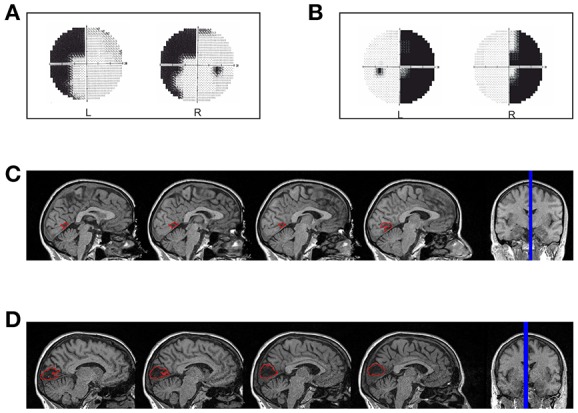
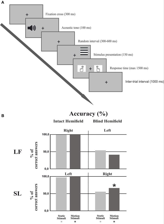
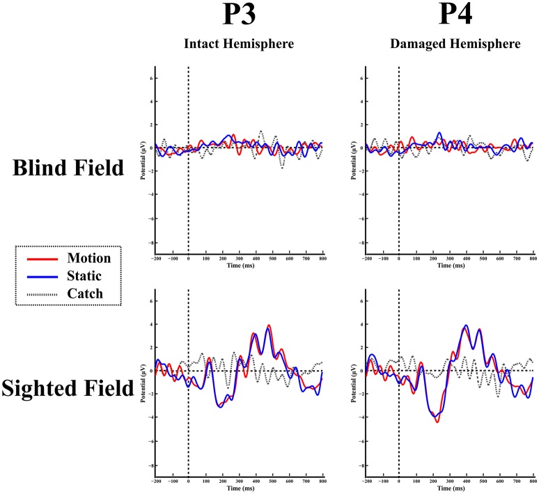
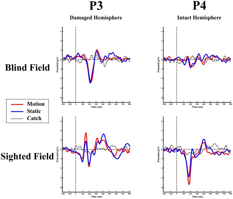
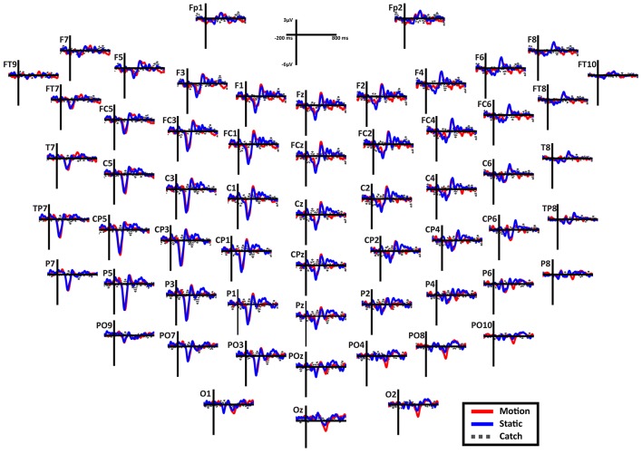
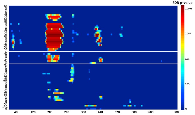
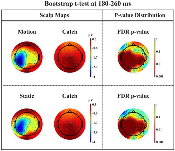
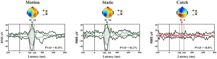
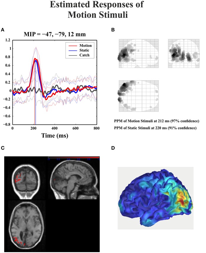
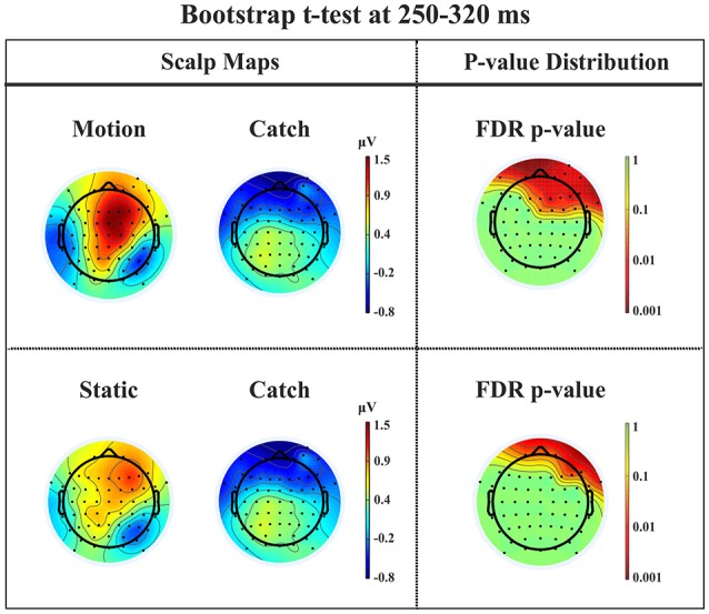
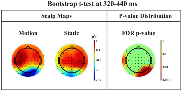
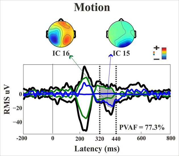
Here we present evidence that a hemianopic patient with a lesion of the left primary visual cortex (V1) showed an unconscious above-chance orientation discrimination with moving rather than static visual gratings presented to the blind hemifield. The patient did not report any perceptual experience of the stimulus features except for a feeling that something appeared in the blind hemifield. Interestingly, in the lesioned left hemisphere, following stimulus presentation to the blind hemifield, we found an event-related potential (ERP) N1 component at a post-stimulus onset latency of 180-260 ms and a source generator in the left BA 19. In contrast, we did not find evidence of the early visual components C1 and P1 and of the later component P300. A positive component (P2a) was recorded between 250 and 320 ms after stimulus onset frontally in both hemispheres. Finally, in the time range 320-440 ms there was a negative peak in right posterior electrodes that was present only for the moving condition. In sum, there were two noteworthy results: Behaviorally, we found evidence of above chance unconscious (blindsight) orientation discrimination with moving but not static stimuli. Physiologically, in contrast to previous studies, we found reliable ERP components elicited by stimuli presented to the blind hemifield at various electrode locations and latencies that are likely to index either the perceptual report of the patient (N1 and P2a) or, the above-chance unconscious performance with moving stimuli as is the case of the posterior ERP negative component. This late component can be considered as the neural correlate of a kind of blindsight enabling feature discrimination only when stimuli are moving and that is subserved by the intact right hemisphere through interhemispheric transfer.

Keywords: blindsight; event related potential; hemianopia; perceptual awareness.


Figures

Figure 1
Figure 1
Hemianopic Patients: (A) Visual field defect in patient LF; (B) Visual field defect in patient SL; (C) reconstruction of the lesion in patient LF; (D) reconstruction of the lesion in patient SL.
Figure 2
Figure 2
Experimental procedure and Behavioral results. (A) Experimental procedure: First, a fixation cross was presented for 300 ms followed by an acoustic tone lasting 100 ms. After a random interval (300–600 ms) the stimulus was presented for 150 ms. The subject had 1500 ms to respond by pressing a keyboard button. The inter-trial interval lasted 1,000 ms. (B) Behavioral results: Percentage of correct responses in each hemifield for each condition in both patients. The asterisk indicates that the number of correct responses in the motion condition was significantly different from the chance level of 50%.
Figure 3
Figure 3
Healthy participant's grand average ERPs for the motion, static and catch condition as recorded at electrode Pz. The top and bottom inlets show the topography scalp map for each condition at the time window of N1 (bottom) and P300 (top).
Figure 4
Figure 4
Patient LF: ERPs in the blind (top) and sighted field (bottom) as recorded at P3 (left-intact hemisphere) and P4 (right-damaged hemisphere) for the three stimulation condition.
Figure 5
Figure 5
Patient SL: ERPs in the blind (top) and sighted field (bottom) as recorded at P3 (left-damaged hemisphere) and P4 (right-intact hemisphere) for the three stimulation condition.
Figure 6
Figure 6
Patient SL: Scalp distribution of the ERPs when the stimulus is presented in the blind field. Note that the large negative peak (N1) is present in most electrodes in the left (damaged) hemisphere, while the frontal peak (P2a) is present in both hemispheres in the fronto-central electrodes. Finally, in the right posterior electrodes is present a late negativity selective for the motion stimuli.
Figure 7
Figure 7
Patient SL: Raster data resulting from the bootstrap ANOVA across all electrodes and the three conditions of blind field stimulation. Color points represent the p-values after the FDR correction for multiple comparisons. Ordinates: left, electrode sites; right, p-values. Abscissae: post-stimulus onset time (ms).
Figure 8
Figure 8
Patient SL: Percentile Bootstrap re-sampling t-test between stimulus against no-stimulus conditions in the time window of the N1. On the left side, Scalp maps representing motion against catch condition (upper panel) and static against catch condition (lower panel); Right side, distribution of the p-values after correction for multiple comparison.
Figure 9
Figure 9
Patient SL: Envelope of the ERP (black line) for the three conditions of stimulus presentation to the blind field. The green line represents the contribution of the most prominent IC when the stimulus, either static or moving is presented. The red line represents the contribution of the most prominent IC during catch trials. The dotted lines represent the time window considered for the PVAF.
Figure 10
Figure 10
Patient SL: 3D source reconstruction of the ERPs when the stimulus was presented in the blind hemifield. (A) Time course of the region with maximal activity for the three conditions. For both motion and static stimuli the MIP is at the same time (corresponding to N1 latency) in the extra-striate cortex (BA19). (B) MIP of the 512 greatest source strengths within MNI space projected onto a glass brain for the motion condition. The area at the highest density correspond to left BA 19. (C) MIP of the statistical map for the motion condition projected on the T1-weighted images of patient SL showing both the lesion and in red the source reconstruction. (D) Summary power image from source reconstruction of motion stimuli presentation to the blind hemifield on a 3D rendered image.
Figure 11
Figure 11
Patient SL: Percentile Bootstrap re-sampling t-test between stimulus against no-stimulus conditions in the time window of the P2a. On the left side, Scalp maps representing the motion against catch conditions (upper panel) and static against catch conditions (lower panel); Right side, distribution of the p-values after correction for multiple comparison.
Figure 12
Figure 12
Patient SL: Percentile Bootstrap re-sampling t-test between Blind Motion and Blind Static conditions in the time window from 320 to 440 ms. On the left side, Scalp maps representing the Bootstrap t-test to compare the motion against static; on the right side the distribution of the p-values after the correction for multiple comparison.
Figure 13
Figure 13
Patient SL: Envelope of the ERP (black line) for the motion condition of stimulus presentation to the blind field. The green and blue lines represent the contribution of the two most prominent ICs. The dotted lines represent the time window considered for the PVAF.

Similar articles

Cited by

References

    1. Ajina S., Bridge H. (2016). Blindsight and unconscious vision: what they teach us about the human visual system. Neuroscientist. 23, pii: 1073858416673817. 10.1177/1073858416673817 - DOI - PMC - PubMed
    1. Ajina S., Kennard C., Rees G., Bridge H. (2015a). Motion area V5/MT+ response to global motion in the absence of V1 resembles early visual cortex. Brain 138, 164–178. 10.1093/brain/awu328 - DOI - PMC - PubMed
    1. Ajina S., Pestilli F., Rokem A., Kennard C., Bridge H. (2015b). Human blindsight is mediated by an intact geniculo-extrastriate pathway. Elife 4:pii: e08935. 10.7554/eLife.08935 - DOI - PMC - PubMed
    1. Ajina S., Rees G., Kennard C., Bridge H. (2015c). Abnormal contrast responses in the extrastriate cortex of blindsight patients. J. Neurosci. 35, 8201–8213. 10.1523/JNEUROSCI.3075-14.2015 - DOI - PMC - PubMed
    1. Azzopardi P., Cowey A. (2001). Motion discrimination in cortically blind patients. Brain. 124, 30–46. 10.1093/brain/124.1.30 - DOI - PubMed

LinkOut - more resources