. 2017 May 23:10:142.
doi: 10.3389/fnmol.2017.00142. eCollection 2017.

Long-Term Assessment of AAV-Mediated Zinc Finger Nuclease Expression in the Mouse Brain

Affiliations

Long-Term Assessment of AAV-Mediated Zinc Finger Nuclease Expression in the Mouse Brain

Muzna Zahur et al. Front Mol Neurosci. .

Abstract

Gene editing tools like TALENs, ZFNs and Crispr/Cas now offer unprecedented opportunities for targeted genetic manipulations in virtually all species. Most of the recent research in this area has concentrated on manipulation of the genome in isolated cells, which then give rise to transgenic animals or modified stem cell lines. Much less is known about applicability of genetic scissors in terminally differentiated, non-dividing cells like neurons of the adult brain. We addressed this question by expression of a pair of ZFNs targeting the murine cathepsin D gene in CNS neurons by means of an optimized AAV viral vector. We show that ZFN expression resulted in substantial depletion of cathepsin D from neuronal lysosomes, demonstrating a robust gene deletion. Importantly, long-term ZFN expression in CNS neurons did not impair essential neuronal functionality and did not cause inflammation or neurodegeneration, suggesting that potent genetic scissors can be expressed safely in the mouse brain. This finding opens up new venues to create novel research models for neurodegenerative disorders.

Keywords: adeno-associated virus; cathepsin D; genome editing; in vivo; zinc finger nuclease.


Figures

FIGURE 1
FIGURE 1
Targeting of the CatD gene in cultured neurons by a 2-vector AAV delivery system. (A) The + strand ZFN (ZFN1) and the – strand ZFN (ZFN2) are expressed from two separate vectors in different layouts: ZFN-WP(D), with hSyn promoter and WPRE; ZFN-P(D), with hSyn promoter but without WPRE; ZFN-NoP(D), with left ITR as promoter; ITR, inverted terminal repeat; WPRE, woodchuck hepatitis virus post-transcriptional control element; bGH-pA, bovine growth hormone polyadenylation site; D, double vector system. (B) Schematic illustrating of ZFN binding strategy to Cathepsin D locus. (C) Western blot analysis of AAV expressed flag-tagged ZFN using cell lysates from mouse cortical neurons at 21 day post-transduction. AAV-EGFP was used as transduction control. (D) Quantitative evaluation of band intensities of western blot analysis reveals more reduction of CatD protein levels by higher levels of ZFN expression. Data given as means from three independent experiments ±SEM.
FIGURE 2
FIGURE 2
Targeting of the CatD gene in cultured neurons by a 1-vector AAV delivery system. (A) The + strand ZFN (ZFN1) and the – strand ZFN (ZFN2) are expressed from a single vector, each by its own hSyn promoter but without WPRE. ITR, inverted terminal repeat; WPRE, woodchuck hepatitis virus post-transcriptional control element; bGH-pA, bovine growth hormone polyadenylation site; S, single vector system; W, week. (B) Western blot analysis of AAV expressed flag-tagged ZFN using cell lysates from mouse cortical neurons at 7 or 21 days post-transduction. AAV-EGFP was used as transduction control. (C) Quantitative evaluation of band intensities of western blot analysis reveals that significant reduction of CatD levels was achieved at 21 days after transduction. Data given as means from three independent experiments ±SEM.
FIGURE 3
FIGURE 3
Frequency and pattern of gene modifications following ZFN administration in mouse neurons. (A) On-target and off-target indel frequencies for the CatD gene after 2 and 3 weeks of ZFN expression. Amplicons for the target sequence of CatD were obtained from the DNA isolated from the neurons transduced with ZFN against CatD. Neurons transduced with the ZFN1 and ZFN2 individually and those with non-CatD ZFN served as negative control. Off-target amplicons were obtained from the DNA collected from the neurons expressing CatD ZFN. (B) Distribution of indel length (Number of deletions and insertions) in CatD gene after 2 and 3 weeks of ZFN expression. For (A,B), error bars reflect SEM from three biological replicates performed on independent experiments. (C) Representative examples of genomic DNA sequences at CatD locus that are modified following ZFN transduction. The unmodified genomic site is the first sequence, followed by the most abundant sequences containing deletions and insertions. The stars represent gaps, a dot for a match on the same strand.
FIGURE 4
FIGURE 4
Zinc finger nuclease expression in primary neurons does not affect essential neuronal functionality. Neurons co-transduced with 1-vector ZFN construct and GCaMP3.5 were subjected to field stimulation (FS). (A) Typical traces obtained from neurons at 7 or 14 days post-transduction are shown, with black horizontal bars representing the 5 s stimulus at 10 Hz. (B) The fractions of excitable neurons were not different between ZFN-expressing neurons or controls. (C) The maximum uptake of calcium upon FS was not different between ZFN expressing neurons and controls. (D) Calcium decay times after FS were not different between ZFN expressing neurons and controls. (E) The number of neurons per mm2 were not different between ZFN expressing neurons and controls.
FIGURE 5
FIGURE 5
Targeting of the CatD gene in striatal neurons of the adult mouse brain by a 1-vector AAV delivery system. (A) Overview of coronal sections showing flag-tagged ZFN (green) expression in the striatum. (B) Striatal tissue sections were prepared at 4, 8, and 16 weeks after the injection of single AAV1/2-ZFN vector and were immunohistochemically stained for CatD (red), the Flag-tag fused to each ZFN (Green; upper two rows of panels) and CatD (red) and DAPI (blue) in higher magnifications (lower row of panels). Images were recorded on a confocal microscope. Scale Bar = 100 μm for the upper two panels and 5 μm for the lower row of panels. (C) Quantification of Cat-D positive lysosomes in transduced and untransduced sides of the striatal area. ∗∗∗Indicates p < 0.0001 Student’s t-test; error bars = SEM.
FIGURE 6
FIGURE 6
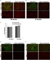
Long-term ZFN expression in the adult mouse brain is safe. (A) Mouse striatal neurons were stained for the neuronal marker NeuN (red) and the ZFN-fused Flag-tag (green) after 8 weeks (A) or 16 weeks (B) of CatD-ZFN expression from the one-vector AAV. Stereological counting revealed that at both times the number of neurons is not different in AAV-CatD-ZFN transduced hemisphere versus the contralateral control hemisphere (C). Brain sections were also stained for the astroglial antigen glial fibrillary acidic protein (GFAP, red) as shown in (D) and for the microglial antigen Iba1 (red) in (E). No evidence for astroglial or microglial activation was detectable in AAV-CatD-ZFN transduced hemisphere as compared to the contralateral control hemisphere. Scale bar = 20 μm.

Similar articles

Cited by

References

    1. Anguela X. M., Sharma R., Doyon Y., Miller J. C., Li H., Haurigot V., et al. (2013). Robust ZFN-mediated genome editing in adult hemophilic mice. Blood 122 3283–3287. 10.1182/blood-2013-04-497354 - DOI - PMC - PubMed
    1. Benes P., Vetvicka V., Fusek M. (2008). Cathepsin D–many functions of one aspartic protease. Crit. Rev. Oncol. Hematol. 68 12–28. 10.1016/j.critrevonc.2008.02.008 - DOI - PMC - PubMed
    1. Carroll D. (2011). Genome engineering with zinc-finger nucleases. Genetics 188 773–782. 10.1534/genetics.111.131433 - DOI - PMC - PubMed
    1. Cradick T. J., Ambrosini G., Iseli C., Bucher P., McCaffrey A. P. (2011). ZFN-site searches genomes for zinc finger nuclease target sites and off-target sites. BMC Bioinformatics 12:152 10.1186/1471-2105-12-152 - DOI - PMC - PubMed
    1. Cullen V., Lindfors M., Ng J., Paetau A., Swinton E., Kolodziej P., et al. (2009). Cathepsin D expression level affects alpha-synuclein processing, aggregation, and toxicity in vivo. Mol. Brain 2:5 10.1186/1756-6606-2-5 - DOI - PMC - PubMed