

Lower Activation in Frontal Cortex and Posterior Cingulate Cortex Observed during Sex Determination Test in Early-Stage Dementia of the Alzheimer Type
- PMID: 28588478
- PMCID: PMC5438965
- DOI: 10.3389/fnagi.2017.00156
Lower Activation in Frontal Cortex and Posterior Cingulate Cortex Observed during Sex Determination Test in Early-Stage Dementia of the Alzheimer Type
Abstract
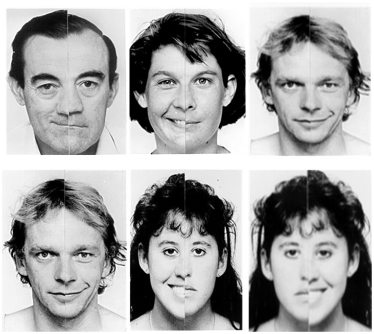
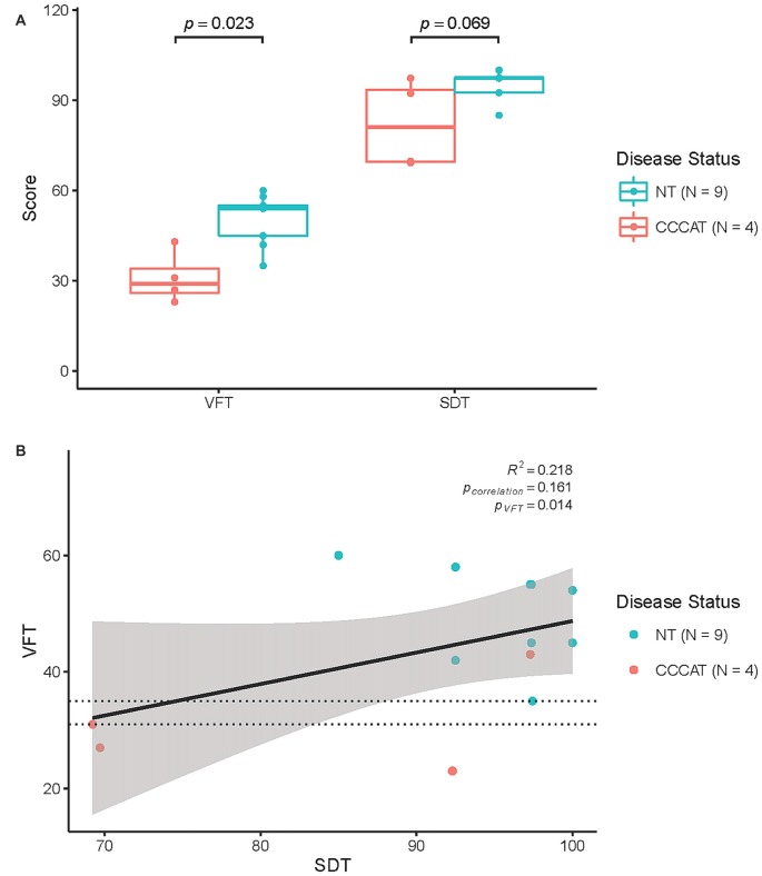
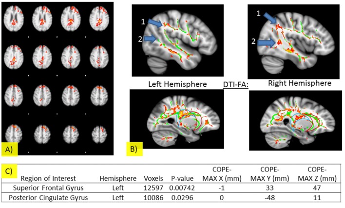
Face-labeling refers to the ability to classify faces into social categories. This plays a critical role in human interaction as it serves to define concepts of socially acceptable interpersonal behavior. The purpose of the current study was to characterize, what, if any, impairments in face-labeling are detectable in participants with early-stage clinically diagnosed dementia of the Alzheimer type (CDDAT) through the use of the sex determination test (SDT). In the current study, four (1 female, 3 males) CDDAT and nine (4 females, 5 males) age-matched neurotypicals (NT) completed the SDT using chimeric faces while undergoing BOLD fMRI. It was expected that CDDAT participants would have poor verbal fluency, which would correspond to poor performance on the SDT. This could be explained by decreased activation and connectivity patterns within the fusiform face area (FFA) and anterior cingulate cortex (ACC). DTI was also performed to test the association of pathological deterioration of connectivity in the uncinate fasciculus (UF) and verbally-mediated performance. CDDAT showed lower verbal fluency test (VFT) performance, but VFT was not significantly correlated to SDT and no significant difference was seen between CDDAT and NT for SDT performance as half of the CDDAT performed substantially worse than NT while the other half performed similarly. BOLD fMRI of SDT displayed differences in the left superior frontal gyrus and posterior cingulate cortex (PCC), but not the FFA or ACC. Furthermore, although DTI showed deterioration of the right inferior and superior longitudinal fasciculi, as well as the PCC, it did not demonstrate significant deterioration of UF tracts. Taken together, early-stage CDDAT may represent a common emerging point for the loss of face labeling ability.
Keywords: Alzheimer; brain networks; chimeric faces; face-processing; neurodegeneration; neuroimaging.
Figures
Similar articles
-
White Matter Deterioration May Foreshadow Impairment of Emotional Valence Determination in Early-Stage Dementia of the Alzheimer Type.Front Aging Neurosci. 2017 Mar 1;9:37. doi: 10.3389/fnagi.2017.00037. eCollection 2017. Front Aging Neurosci. 2017. PMID: 28298891 Free PMC article.
-
ACC and IPL networks in the perception of the faces of parents during selective tasks.Brain Imaging Behav. 2016 Dec;10(4):1172-1183. doi: 10.1007/s11682-015-9486-1. Brain Imaging Behav. 2016. PMID: 26613720
-
Functional connectivity of the fusiform gyrus during a face-matching task in subjects with mild cognitive impairment.Brain. 2006 May;129(Pt 5):1113-24. doi: 10.1093/brain/awl051. Epub 2006 Mar 6. Brain. 2006. PMID: 16520329
-
Sex and performance level effects on brain activation during a verbal fluency task: a functional magnetic resonance imaging study.Cortex. 2009 Feb;45(2):164-76. doi: 10.1016/j.cortex.2007.09.006. Epub 2008 Feb 5. Cortex. 2009. PMID: 19150518
-
The structural and functional connectivity of the posterior cingulate cortex: comparison between deterministic and probabilistic tractography for the investigation of structure-function relationships.Neuroimage. 2014 Nov 15;102 Pt 1:118-27. doi: 10.1016/j.neuroimage.2013.12.022. Epub 2013 Dec 21. Neuroimage. 2014. PMID: 24365673 Review.
Cited by
-
Facial expression recognition deficits in frontotemporal dementia and Alzheimer's disease: a meta-analytic investigation of effects of phenotypic variant, task modality, geographical region and symptomatic specificity.J Neurol. 2023 Dec;270(12):5731-5755. doi: 10.1007/s00415-023-11927-4. Epub 2023 Sep 6. J Neurol. 2023. PMID: 37672106 Review.
References
-
- Albert M. S., DeKosky S. T., Dickson D., Dubois B., Feldman H. H., Fox N. C., et al. . (2011). The diagnosis of mild cognitive impairment due to Alzheimer disease: recommendations from the national institute on aging-Alzheimer’s association workgroups on diagnostic guidelines for Alzheimer disease. Alzheimers Dement. 7, 270–279. 10.1016/j.jalz.2011.03.008 - DOI - PMC - PubMed
-
- Bartzokis G., Sultzer D., Lu P. H., Nuechterlein K. H., Mintz J., Cummings J. L. (2004). Heterogenous age related breakdown of white matter structural integrity: implications for cortical “disconnection” in aging and Alzheimer disease. Neurobiol. Aging 25, 843–851. 10.1016/j.neurobiolaging.2003.09.005 - DOI - PubMed
Grants and funding
LinkOut - more resources
Full Text Sources
Other Literature Sources
Research Materials