

Case Reports
doi: 10.1002/ccr3.952.
eCollection 2017 Jun.
Bilateral hydronephrosis due to obstructive ureteral stone associated with norovirus gastroenteritis
Affiliations
- PMID: 28588843
- PMCID: PMC5458008
- DOI: 10.1002/ccr3.952
Item in Clipboard
Case Reports
Bilateral hydronephrosis due to obstructive ureteral stone associated with norovirus gastroenteritis
Clin Case Rep.
.
Abstract
Recently, cases of urinary tract calculi causing hydronephrosis and postrenal renal failure associated with viral gastroenteritis were documented, yet few were related to norovirus. During norovirus gastroenteritis, observation of oliguria, aciduria, low FENa value, and elevation of blood or urinary uric acid level may necessitate clinical workout for nephrolithiasis.
Keywords: Gastroenteritis; hydronephrosis; norovirus; ureteral calculi; uric acid.
Figures
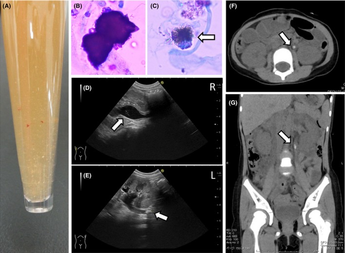
(A) Urine appearance. White and brown fine sandy stones were observed in urine. (B) Ammonium acid urate crystal with haw‐like appearance in the urinary sediment (Sternheimer stain). (C) Sodium urate crystal in the urinary sediment (Sternheimer stain) with spinal‐like appearance (arrows). (D, E) Abdominal ultrasound revealing dilated bilateral pelvises and calculi in the left ureter on admission. Arrows show some fine high echoic lesions in bilateral pelvises. (F, G) Abdominal computed tomography showing dilated calculi in the left ureter on admission. (F) Axial section and (G) coronal section. Arrows indicate urinary stone (4.2 × 8.7 mm) in the left upper ureter.
Similar articles
-
Acute kidney injury due to ammonium acid urate stones in a patient with adenovirus gastroenteritis: a case report.BMC Urol. 2022 Jan 15;22(1):5. doi: 10.1186/s12894-022-00954-4. BMC Urol. 2022. PMID: 35033051 Free PMC article.
-
[Acute renal failure due to obstructive ureteral stone associated with norovirus gastroenteritis in an infant with congenital solitary kidney].Nihon Hinyokika Gakkai Zasshi. 2014 Oct;105(4):224-8. doi: 10.5980/jpnjurol.105.224. Nihon Hinyokika Gakkai Zasshi. 2014. PMID: 25757355 Japanese.
-
[Two cases of infants with acute renal failure due to bilateral obstructive ureteral stones associated with rotavirus gastroenteritis].Nihon Hinyokika Gakkai Zasshi. 2010 Jan;101(1):29-33. doi: 10.5980/jpnjurol.101.29. Nihon Hinyokika Gakkai Zasshi. 2010. PMID: 20158076 Japanese.
-
The bladder ran dry: bilateral ureteral obstruction.BMJ Case Rep. 2017 Aug 7;2017:bcr2016218173. doi: 10.1136/bcr-2016-218173. BMJ Case Rep. 2017. PMID: 28784872 Free PMC article. Review.
-
[Radiotransparent lithiasis. Diagnosis and treatment].Arch Esp Urol. 2001 Nov;54(9):997-1008. Arch Esp Urol. 2001. PMID: 11789377 Review. Spanish.
Cited by
-
Traditional Chinese medicine on treating ureteral calculi: A systematic review and meta-analysis protocol.Medicine (Baltimore). 2019 Sep;98(37):e17057. doi: 10.1097/MD.0000000000017057. Medicine (Baltimore). 2019. PMID: 31517825 Free PMC article.
-
Acute kidney injury due to ammonium acid urate stones in a patient with adenovirus gastroenteritis: a case report.BMC Urol. 2022 Jan 15;22(1):5. doi: 10.1186/s12894-022-00954-4. BMC Urol. 2022. PMID: 35033051 Free PMC article.
-
Acute renal failure due to severe bilateral ureteropelvic junction obstruction treated by urinary drainage in a 2-year-old infant.IJU Case Rep. 2021 Nov 11;5(1):70-73. doi: 10.1002/iju5.12397. eCollection 2022 Jan. IJU Case Rep. 2021. PMID: 35005479 Free PMC article.
-
Paediatrics: how to manage viral gastroenteritis.Drugs Context. 2021 Mar 26;10:2020-11-7. doi: 10.7573/dic.2020-11-7. eCollection 2021. Drugs Context. 2021. PMID: 33828604 Free PMC article. Review.
References
-
- Gearhart, J. P. , Herzberg G. Z., and Jeffs R. D.. 1991. Childhood urolithiasis: experiences and advances. Pediatrics 87:445–450. - PubMed
-
- Sarkissian, A. , Babloyan A., Arikyants N., Hesse A., Blau N., and Leumann E.. 2001. Pediatric urolithiasis in Armenia: a study of 198 patients observed from 1991 to 1999. Pediatr. Nephrol. 16:728–732. - PubMed
-
- Shirasu, A. , Ashida A., Matsumura H., Nakakura H., and Tamai H.. 2015. Clinical characteristics of rotavirus gastroenteritis with urinary crystals. Pediatr. Int. 57:917–921. - PubMed
-
- Morita, T. , Ashida A., Fujieda M., Hayashi A., Maeda A., Ohta K., et al. 2010. Four cases of postrenal renal failure induced by renal stone associated with rotavirus infection. Clin. Nephrol. 73:398–402. - PubMed
Publication types
LinkOut - more resources
Full Text Sources
Other Literature Sources
