Case Reports
. 2017 Apr 25:7:17.
doi: 10.4103/jcis.JCIS_96_16. eCollection 2017.

Vaginal Ewing Sarcoma: An Uncommon Clinical Entity in Pediatric Patients

Affiliations
Case Reports

Vaginal Ewing Sarcoma: An Uncommon Clinical Entity in Pediatric Patients

Nathan M Cross et al. J Clin Imaging Sci. .

Abstract

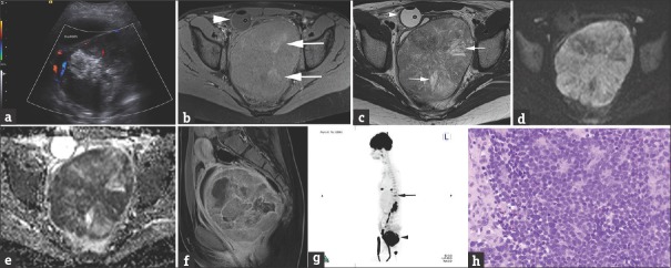
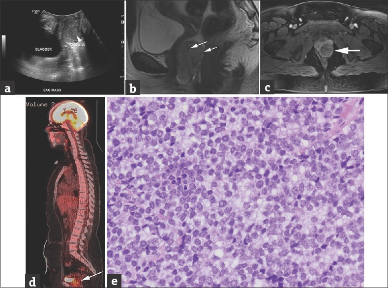
Ewing sarcoma, including classical Ewing sarcoma of the bone and primitive neuroectodermal tumors arising in bone or extraosseous primary sites, is a highly aggressive childhood neoplasm. We present two cases of Ewing sarcoma arising from the vagina in young girls. Previously reported cases in literature focused on their pathologic rather than radiographic features. We describe the spectrum of multimodality imaging appearances of Ewing sarcoma at this unusual primary site. Awareness of vaginal Ewing tumors may facilitate prompt diagnosis and lead to a different surgical approach than the more commonly encountered vaginal rhabdomyosarcoma.

Keywords: Ewing sarcoma; pediatric; vaginal.


Conflict of interest statement

There are no conflicts of interest.

Figures

Figure 1
Figure 1
A 12-year-old female presented with acute onset of urinary retention. (a) Transverse, color flow ultrasound image through the pelvis demonstrates a large, heterogeneous mass posterior to the bladder measuring 10.1 cm × 8.8 cm × 12.4 cm (anteroposterior × transverse × craniocaudal), with minimal vascularity, and large hyperechoic foci, some of which shadow, suggestive of calcifications. (b) Axial T1 fat-sat precontrast MRI with a Foley decompressing the bladder (arrowhead) better depicts the large heterogeneous pelvic mass, containing areas of high T1 signal (arrows) corresponding to calcifications present on low-dose noncontrast CT for attenuation correction performed as part of the 18F FDG-PET (not shown). (c) Axial T2 image at a similar level as b shows areas of high T2 signal representing cystic necrosis. (d and e) Axial diffusion and apparent diffusion coefficient map images show extensive restrictive diffusion. (f) Sagittal contrast-enhanced T1 MRI of the pelvis confirms that the large, heterogeneously enhancing mass with areas of necrosis arises from the posterior wall of the vagina, compresses the bladder and the rectum and displaces the uterus superiorly. (g) Sagittal three-dimensional maximum intensity projection 18F FDG-PET/CT demonstrates heterogeneous hypermetabolic uptake in the pelvic mass (arrowhead) as well as additional hypermetabolic foci in the lung bases and at the T11 vertebral body (arrow), consistent with metastatic lesions. (h) Histologic examination shows sheets of small round blue cells with inconspicuous nucleoli (H and E, ×400). The cells are focally arranged in primitive rosettes (top center).
Figure 2
Figure 2
A 15-year-old female presented with progressive abdominal discomfort, profuse menstrual bleeding, and vaginal discharge. (a) Grayscale sagittal ultrasound image through the pelvis obtained at initial presentation demonstrate a slightly heterogeneous, predominantly isoechoic, 3.6 cm × 3.6 cm × 5.9 cm (anteroposterior x transverse x craniocaudal), vaginal mass (arrowhead on a). (b and c) Sagittal T2 (b) and axial postcontrast T1 MRI (c) show the lesion arising from the left vaginal sidewall demonstrating intermediate to high signal on T2-weighted images with few serpentine flow voids (arrows on b) and intermediate to low T1 signal, with heterogeneous contrast enhancement on (c). (d) Sagittal fused 18F FDG-PET/CT demonstrates increased FDG uptake in the mass (arrow) posterior to the bladder. (e) Histologic examination shows sheets of small round blue cells with inconspicuous nucleoli and a suggestion of rosette formation (H and E, ×400 magnification). Pathology image courtesy of Dr. Aashiyana Koreishi, Puget Sound Institute of Pathology - Franciscan Health System.

References

    1. de Alava E, Lessnick SL, Sorensen PH. WHO Classification of Tumours of Soft Tissue and Bone. Lyon: IARC Press; 2013. Ewing sarcoma; pp. 305–9.
    1. Juergens H, Daw NC, Geoerger B, Ferrari S, Villarroel M, Aerts I, et al. Preliminary efficacy of the anti-insulin-like growth factor type 1 receptor antibody figitumumab in patients with refractory Ewing sarcoma. J Clin Oncol. 2011;29:4534–40. - PMC - PubMed
    1. Murphey MD, Senchak LT, Mambalam PK, Logie CI, Klassen-Fischer MK, Kransdorf MJ. From the radiologic pathology archives: Ewing sarcoma family of tumors: Radiologic-pathologic correlation. Radiographics. 2013;33:803–31. - PubMed
    1. Applebaum MA, Worch J, Matthay KK, Goldsby R, Neuhaus J, West DC, et al. Clinical features and outcomes in patients with extraskeletal Ewing sarcoma. Cancer. 2011;117:3027–32. - PMC - PubMed
    1. Bancalari E, de Álava E, Tardío JC. Primary vaginal Ewing sarcoma: Case report and review of the literature. Int J Surg Pathol. 2012;20:305–10. - PubMed

Publication types