Review
. 2017:2017:7897325.
doi: 10.1155/2017/7897325. Epub 2017 May 15.

Skeletal Muscle and Lymphocyte Mitochondrial Dysfunctions in Septic Shock Trigger ICU-Acquired Weakness and Sepsis-Induced Immunoparalysis

Affiliations
Review

Skeletal Muscle and Lymphocyte Mitochondrial Dysfunctions in Septic Shock Trigger ICU-Acquired Weakness and Sepsis-Induced Immunoparalysis

Quentin Maestraggi et al. Biomed Res Int. 2017.

Abstract

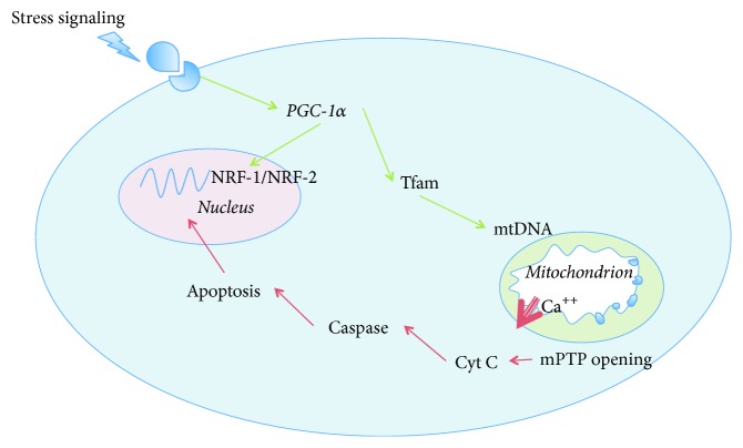
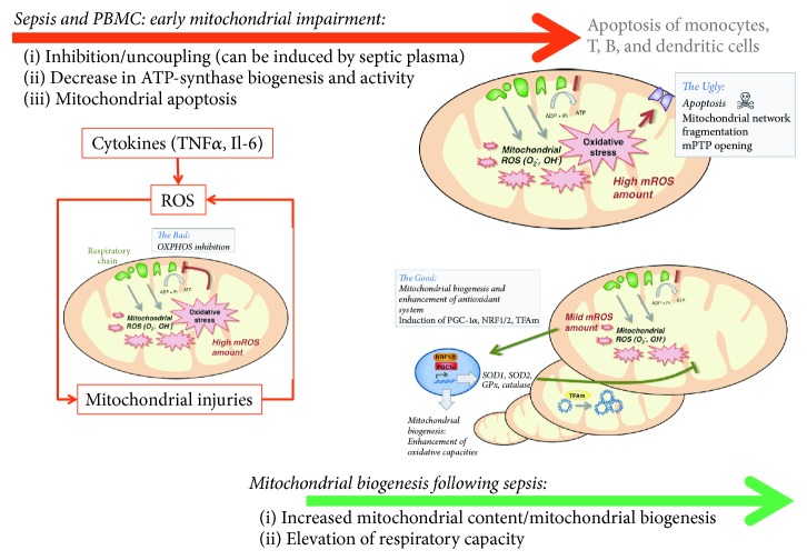
Fundamental events driving the pathological processes of septic shock-induced multiorgan failure (MOF) at the cellular and subcellular levels remain debated. Emerging data implicate mitochondrial dysfunction as a critical factor in the pathogenesis of sepsis-associated MOF. If macrocirculatory and microcirculatory dysfunctions undoubtedly participate in organ dysfunction at the early stage of septic shock, an intrinsic bioenergetic failure, sometimes called "cytopathic hypoxia," perpetuates cellular dysfunction. Short-term failure of vital organs immediately threatens patient survival but long-term recovery is also severely hindered by persistent dysfunction of organs traditionally described as nonvital, such as skeletal muscle and peripheral blood mononuclear cells (PBMCs). In this review, we will stress how and why a persistent mitochondrial dysfunction in skeletal muscles and PBMC could impair survival in patients who overcome the first acute phase of their septic episode. First, muscle wasting protracts weaning from mechanical ventilation, increases the risk of mechanical ventilator-associated pneumonia, and creates a state of ICU-acquired muscle weakness, compelling the patient to bed. Second, failure of the immune system ("immunoparalysis") translates into its inability to clear infectious foci and predisposes the patient to recurrent nosocomial infections. We will finally emphasize how mitochondrial-targeted therapies could represent a realistic strategy to promote long-term recovery after sepsis.


Figures

Figure 1
Figure 1
Succession of events leading to sepsis-induced mitochondrial dysfunction. (1) Increased activity of inducible NO-synthase. (2) Increased mitochondrial superoxide anion generation. (3) Production of peroxynitrite. (4) Nitrosylation of respiratory chain complexes. (5) Decreased membrane potential. (6) Opening of mitochondrial transition pore. Greek numbers refer to mitochondrial complexes. iNOS: inducible nitric oxide synthase; NO: nitric oxide; O2: superoxide anion; ONOO: peroxynitrite; O2: oxygen; ADP: adenosine diphosphate; ATP: adenosine triphosphate; H+: proton; FADH2: reduced flavin-adenine dinucleotide; FAD+: oxidized flavin-adenine dinucleotide; NADH: reduced nicotinamide adenine dinucleotide; NAD+: oxidized nicotinamide adenine dinucleotide; Ca++: ionized calcium; mPTP: mitochondrial permeability transition pore.
Figure 2
Figure 2
Nucleus-mitochondria crosstalk for apoptosis and mitochondrial hormesis induction: key role of PGC-1α, Tfam, and NRF-1/2. (i) Activation of caspase-3 and caspase-7 after mPTP opening leading to apoptosis. (ii) Activation of mitochondrial hormesis in response to stress stimulation (i.e., β-adrenergic). PGC-1α plays a pivotal role for transcription of both nuclear and mitochondrial genes leading to increased mitohormesis. PGC-1α: peroxisome proliferator-activated receptor-γ coactivator 1α; NRF-1/2: nuclear respiratory factor 1/2; Tfam: mitochondrial transcription factor A; mtDNA: mitochondrial desoxyribonucleic acid; Ca++: ionized calcium; mPTP: mitochondrial permeability transition pore; Cyt c: cytochrome c.
Figure 3
Figure 3
Time course of mitochondrial impairment and mitochondrial biogenesis following sepsis in peripheral blood mononuclear cells (PBMCs). TNFα: tumor necrosis factor α; Il-6: interleukin-6; ROS: reactive oxygen species; SOD: superoxide dismutase, GPx: glutathione peroxidase; NRF-1/2: nuclear respiratory factor 1/2; PGC-1α: peroxisome proliferator-activated receptor-γ coactivator 1α; Tfam: mitochondrial transcription factor A. Adapted from Lejay et al. with permission [27].

References

    1. Singer M., Deutschman C. S., Seymour C. W., et al. The third international consensus definitions for sepsis and septic shock (sepsis-3) The Journal of the American Medical Association. 2016;315(8):801–810. doi: 10.1001/jama.2016.0287. - DOI - PMC - PubMed
    1. Bone R. C., Balk R. A., Cerra F. B., et al. Definitions for sepsis and organ failure and guidelines for the use of innovative therapies in sepsis. The ACCP/SCCM Consensus Conference Committee. American College of Chest Physicians/Society of Critical Care Medicine. Chest. 1992;101(6):1644–1655. doi: 10.1378/chest.101.6.1644. - DOI - PubMed
    1. Levy M. M., Fink M. P., Marshall J. C., et al. 2001 SCCM/ESICM/ACCP/ATS/SIS international sepsis definitions conference. Critical Care Medicine. 2003;31(4):1250–1256. doi: 10.1097/01.CCM.0000050454.01978.3B. - DOI - PubMed
    1. Shankar-Hari M., Phillips G. S., Levy M. L., et al. Developing a new definition and assessing new clinical criteria for septic shock. The Journal of the American Medical Association. 2016;315(8):775–787. doi: 10.1001/jama.2016.0289. - DOI - PMC - PubMed
    1. Vincent J.-L., Moreno R., Takala J., et al. The SOFA (Sepsis-related Organ Failure Assessment) score to describe organ dysfunction/failure. Intensive Care Medicine. 1996;22(7):707–710. doi: 10.1007/s001340050156. - DOI - PubMed

MeSH terms