

Age-Specific Prevalence and Clinical Characteristics of Humeral Medial Epicondyle Apophysitis and Osteochondritis Dissecans: Ultrasonographic Assessment of 4249 Players
- PMID: 28589162
- PMCID: PMC5446105
- DOI: 10.1177/2325967117707703
Age-Specific Prevalence and Clinical Characteristics of Humeral Medial Epicondyle Apophysitis and Osteochondritis Dissecans: Ultrasonographic Assessment of 4249 Players
Abstract
Background: Traction apophysitis of medial epicondyle (MEC) lesions and osteochondritis dissecans (OCD) of the capitellum are common elbow injuries in adolescent baseball players. However, the age-specific prevalence of these pathologies and their influence on elbow pain remain unknown.
Purpose: To investigate the age-specific prevalence of each MEC lesion and capitellar OCD and to identify the incidence of elbow pain in each condition.
Study design: Descriptive epidemiology study.
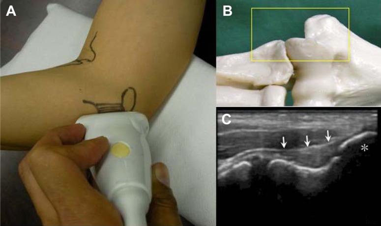
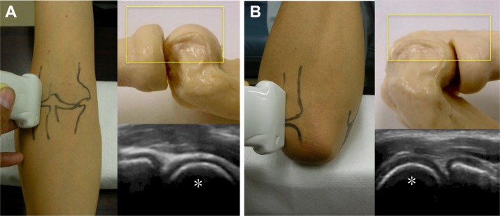
Methods: Study participants consisted of 4249 baseball players aged 6 to 17 years. A questionnaire was used to assess history of elbow pain, and morphological changes of the elbow joint were assessed using ultrasonography.
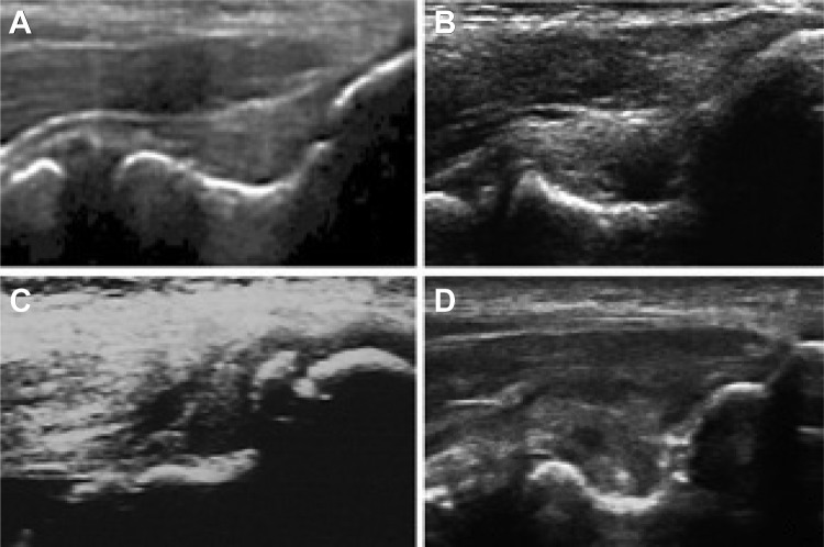
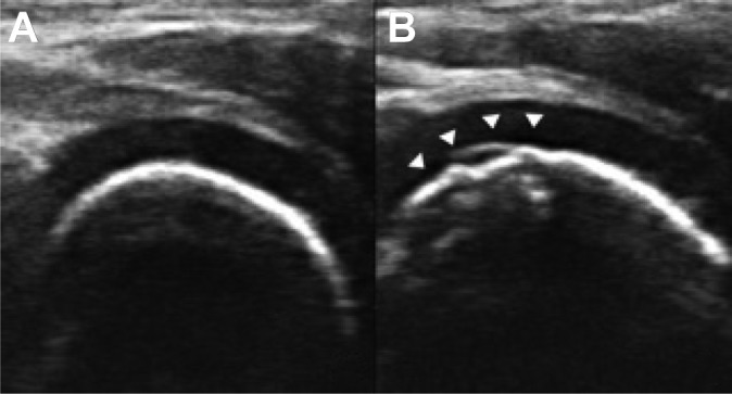
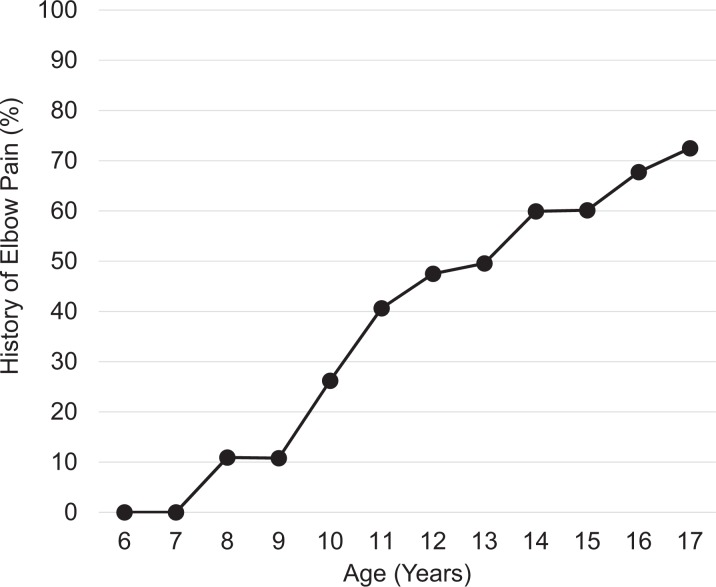
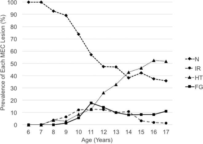
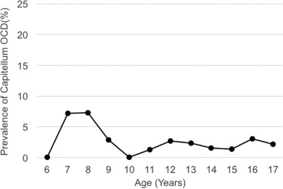
Results: Regarding MEC lesions, fragmented (FG) and irregular (IR) lesions both reached their greatest respective prevalence at 11 to 12 years of age. After 14 years of age, IR decreased sharply, whereas FG was maintained at approximately 10%. Hypertrophic (HT) lesions increased sharply, reaching over 50% at 16 years of age, while there was a decrease in IR and FG lesions in the same age group. The prevalence of capitellar OCD remained the same (approximately 2%) throughout all ages except for in players aged 7 to 8 years (>7%). Players with MEC lesions had significantly greater prevalence of a history of elbow pain compared with those without (68.0% vs 39.1%) and were at a significantly greater risk for FG lesions (odds ratio [OR], 4.04; 95% CI, 3.16-5.22) compared with IR (OR, 3.22; 95% CI, 2.44-4.27) and HT lesions (OR, 2.03; 95% CI, 1.75-2.36). Players with capitellar OCD also had a significantly greater risk of a history of elbow pain (OR, 2.34; 95% CI, 1.40-4.11).
Conclusion: Controlling the amount of practice and its intensity according to the condition of each player in the preadolescent and adolescent periods may be important in accelerating bony healing and decreasing preventable elbow pain in adulthood.
Keywords: age-specific prevalence; elbow pain; medial epicondylar apophysitis; osteochondritis dissecans.
Conflict of interest statement
The authors declared that they have no conflicts of interest in the authorship and publication of this contribution.
Figures
Similar articles
-
Prevalence and Clinical Characteristics of Osteochondritis Dissecans of the Humeral Capitellum Among Adolescent Baseball Players.Am J Sports Med. 2014 Aug;42(8):1963-71. doi: 10.1177/0363546514536843. Epub 2014 Jun 18. Am J Sports Med. 2014. PMID: 24944293
-
Cumulative Incidence of Osteochondritis Dissecans of the Capitellum in Preadolescent Baseball Players.Arthroscopy. 2019 Jan;35(1):60-66. doi: 10.1016/j.arthro.2018.08.034. Arthroscopy. 2019. PMID: 30611367
-
Prevalence of Osteochondritis Dissecans of the Capitellum in Young Baseball Players: Results Based on Ultrasonographic Findings.Orthop J Sports Med. 2014 Aug 12;2(8):2325967114545298. doi: 10.1177/2325967114545298. eCollection 2014 Aug. Orthop J Sports Med. 2014. PMID: 26535356 Free PMC article.
-
Diagnosis and treatment of osteochondritis dissecans of the humeral capitellum.J Orthop Sci. 2018 Mar;23(2):213-219. doi: 10.1016/j.jos.2017.11.013. Epub 2017 Dec 22. J Orthop Sci. 2018. PMID: 29276039 Review.
-
Osteochondritis Dissecans of the Capitellum :Review of the Literature.J Med Invest. 2020;67(3.4):217-221. doi: 10.2152/jmi.67.217. J Med Invest. 2020. PMID: 33148891 Review.
Cited by
-
Normal imaging laterality on magnetic resonance imaging of the medial epicondyle of the elbow on the dominant side of adolescent male baseball players.Skeletal Radiol. 2018 Sep;47(9):1237-1244. doi: 10.1007/s00256-018-2921-9. Epub 2018 Mar 9. Skeletal Radiol. 2018. PMID: 29523906
-
A high rate of children and adolescents return to sport after surgical treatment of osteochondritis dissecans of the elbow: a systematic review and meta-analysis.Knee Surg Sports Traumatol Arthrosc. 2021 Dec;29(12):4041-4066. doi: 10.1007/s00167-021-06489-9. Epub 2021 Feb 23. Knee Surg Sports Traumatol Arthrosc. 2021. PMID: 33620512
-
Deep learning-based osteochondritis dissecans detection in ultrasound images with humeral capitellum localization.Int J Comput Assist Radiol Surg. 2024 Nov;19(11):2143-2152. doi: 10.1007/s11548-023-03040-8. Epub 2024 Jan 17. Int J Comput Assist Radiol Surg. 2024. PMID: 38233599 Free PMC article.
-
Outcomes of Arthroscopic Surgical Treatment of Osteochondral Lesions of the Elbow in Pediatric and Adolescent Athletes.Orthop J Sports Med. 2020 Nov 9;8(11):2325967120963054. doi: 10.1177/2325967120963054. eCollection 2020 Nov. Orthop J Sports Med. 2020. PMID: 33225011 Free PMC article.
-
Osteochondritis dissecans lesions of the capitellum in overhead athletes: a review of current evidence and proposed treatment algorithm.Curr Rev Musculoskelet Med. 2019 Mar;12(1):1-12. doi: 10.1007/s12178-019-09528-8. Curr Rev Musculoskelet Med. 2019. PMID: 30645727 Free PMC article. Review.
References
-
- Baseball Federation of Japan. http://www.baseballjapan.org/jpn/uploaded_data/bfj_news/doc/0125/undoki2.... Published May 15, 2015. Accessed September 15, 2016.
-
- Bauer M, Jonsson K, Josefsson PO, Lindén B. Osteochondritis dissecans of the elbow. A long-term follow-up study. Clin Orthop Relat Res. 1992;284:156–160. - PubMed
-
- Bradley JP, Petrie RS. Osteochondritis dissecans of the humeral capitellum. Diagnosis and treatment. Clin Sports Med. 2001;20:565–590. - PubMed
-
- Brogdon BG, Crow NE. Little Leaguer’s elbow. AJR Am J Roentgenol. 1960;83:671–675. - PubMed
LinkOut - more resources
Full Text Sources
Other Literature Sources
