. 2017 May 9;4(6):369-380.
doi: 10.1002/acn3.418. eCollection 2017 Jun.

CNS Aquaporin-4-specific B cells connect with multiple B-cell compartments in neuromyelitis optica spectrum disorder

Affiliations

CNS Aquaporin-4-specific B cells connect with multiple B-cell compartments in neuromyelitis optica spectrum disorder

Markus C Kowarik et al. Ann Clin Transl Neurol. .

Abstract

Objectives: Neuromyelitis optica spectrum disorder (NMOSD) is a severe inflammatory disorder of the central nervous system (CNS) targeted against aquaporin-4 (AQP4). The origin and trafficking of AQP4-specific B cells in NMOSD remains unknown.

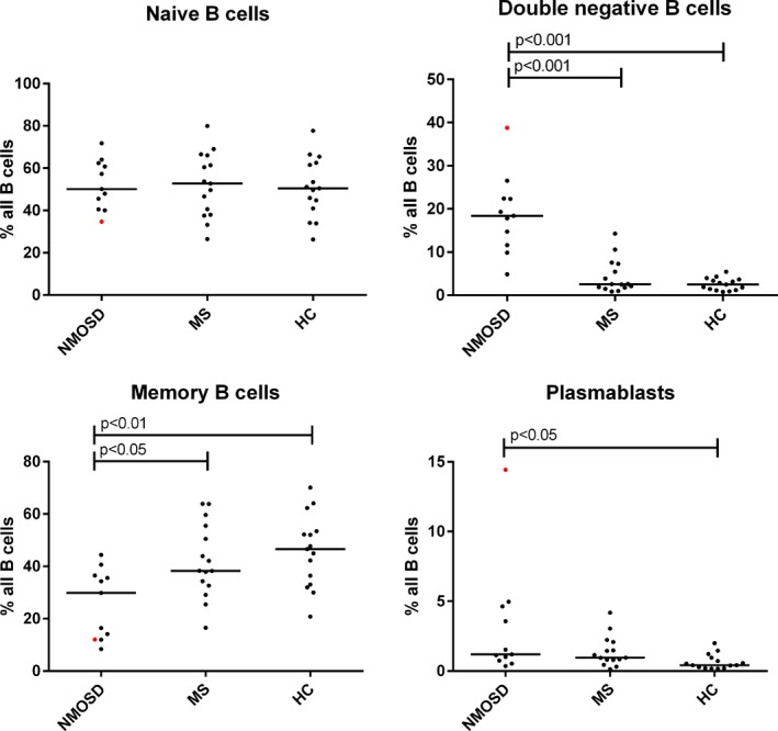
Methods: Peripheral (n = 7) and splenic B cells (n = 1) recovered from seven NMOSD patients were sorted into plasmablasts, naïve, memory, and CD27-IgD- double negative (DN) B cells, and variable heavy chain (VH) transcriptome sequences were generated by deep sequencing. Peripheral blood (PB) VH repertoires were compared to the same patient's single-cell cerebrospinal fluid (CSF) plasmablast (PB) VH transcriptome, CSF immunoglobulin (Ig) proteome, and serum Ig proteome. Recombinant antibodies were generated from paired CSF heavy- and light chains and tested for AQP4 reactivity.

Results: Approximately 9% of the CSF VH sequences aligned with PB memory B cells, DN B cells, and plasmablast VH sequences. AQP4-specific VH sequences were observed in each peripheral B-cell compartment. Lineage analysis of clonally related VH sequences indicates that CSF AQP4-specific B cells are closely related to an expanded population of DN B cells that may undergo antigen-specific B-cell maturation within the CNS. CSF and serum Ig proteomes overlapped with the VH sequences from each B-cell compartment; the majority of matches occurring between the PB VH sequences and serum Ig proteome.

Interpretation: During an acute NMOSD relapse, a dynamic exchange of B cells occurs between the periphery and CNS with AQP4-specific CSF B cells emerging from postgerminal center memory B cells and plasmablasts. Expansion of the PB DN B-cell compartment may be a potential biomarker of NMOSD activity.


Figures

Figure 1
Figure 1
Gating strategy for naïve, CD27 + memory, CD27‐ double negative B cells, and plasmablasts.
Figure 2
Figure 2
Hierarchical maturation diagrams (trees) of related CSF and blood VH sequences. Trees are rooted to germline sequences (GL), and the number of mutations between VH sequences are noted (a single mutation is left blank). Unknown intermediates are symbolized as white circles. (A) The most common representative VH tree showing an unknown intermediate linking a CSF plasmablast to peripheral B cells (12/14 VH Trees). (B) A single peripheral blood B‐cell VH sequence directly links to a clonally related CSF plasmablast (2/14 Ig Trees). (C) CSF plasmablast VH sequence hierarchically linked to a peripheral blood VH sequence (1/14 Ig Trees). GL, Germline; MEM, memory B cell; DN, double negative B cells; PBL, plasmablast, SP PBL, spleen plasmablast.
Figure 3
Figure 3
Percentage distribution of peripheral blood B‐cell populations (naïve, memory, and double negative B cells, plasmablasts) in neuromyelitis spectrum disease (NMOSD), healthy controls (HC), multiple sclerosis (MS) subjects. Red dots indicate NMOSD patient treated with rituximab.
Figure 4
Figure 4
B‐cell trafficking and antibody production in NMOSD. Antigen‐experienced (AQP4‐reactive) B cells migrate between both compartments. While some B cells undergo clonal expansion and affinity maturation in the CNS, most of the AQP4‐specific germinal center reactions take place in the periphery. AQP4‐specific antibodies are produced locally by antibody secreting cells in the CNS; however, a substantial component transits from the serum through an open blood–brain barrier.

References

    1. Bennett JL, Lam C, Kalluri SR, et al. Intrathecal pathogenic anti‐aquaporin‐4 antibodies in early neuromyelitis optica. Ann Neurol 2009;66:617–629. - PMC - PubMed
    1. Lennon VA, Wingerchuk DM, Kryzer TJ, et al. A serum autoantibody marker of neuromyelitis optica: distinction from multiple sclerosis. Lancet 2004;364:2106–2112. - PubMed
    1. Lennon VA, Kryzer TJ, Pittock SJ, et al. IgG marker of optic‐spinal multiple sclerosis binds to the aquaporin‐4 water channel. J Exp Med 2005;202:473–477. - PMC - PubMed
    1. Ratelade J, Zhang H, Saadoun S, et al. Neuromyelitis optica IgG and natural killer cells produce NMO lesions in mice without myelin loss. Acta Neuropathol 2012;123:861–872. - PMC - PubMed
    1. Papadopoulos MC, Bennett JL, Verkman AS. Treatment of neuromyelitis optica: state‐of‐the‐art and emerging therapies. Nat Rev Neurol 2014;10:493–506. - PMC - PubMed

LinkOut - more resources