Review
. 2017 May 16;4(6):422-445.
doi: 10.1002/acn3.419. eCollection 2017 Jun.

Immune and myodegenerative pathomechanisms in inclusion body myositis

Affiliations
Review

Immune and myodegenerative pathomechanisms in inclusion body myositis

Christian W Keller et al. Ann Clin Transl Neurol. .

Abstract

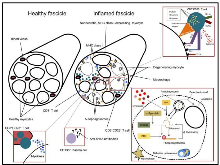
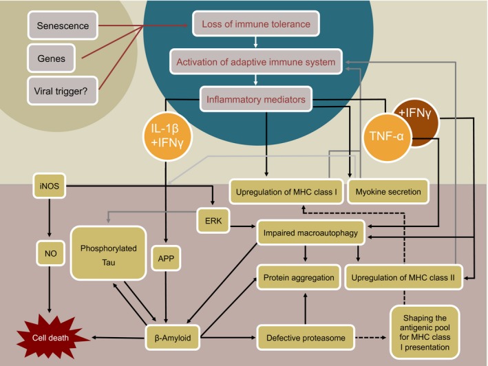
Inclusion Body Myositis (IBM) is a relatively common acquired inflammatory myopathy in patients above 50 years of age. Pathological hallmarks of IBM are intramyofiber protein inclusions and endomysial inflammation, indicating that both myodegenerative and inflammatory mechanisms contribute to its pathogenesis. Impaired protein degradation by the autophagic machinery, which regulates innate and adaptive immune responses, in skeletal muscle fibers has recently been identified as a potential key pathomechanism in IBM. Immunotherapies, which are successfully used for treating other inflammatory myopathies lack efficacy in IBM and so far no effective treatment is available. Thus, a better understanding of the mechanistic pathways underlying progressive muscle weakness and atrophy in IBM is crucial in identifying novel promising targets for therapeutic intervention. Here, we discuss recent insights into the pathomechanistic network of mutually dependent inflammatory and degenerative events during IBM.


Figures

Figure 1
Figure 1
Scheme of the pathological changes in inclusion body myositis compared to healthy muscle. Mainly nonnecrotic, MHC class I‐expressing myofibers are surrounded by invading CD8+ CD28 T cells, which is the predominant immune subset in the endomysial infiltrates. These CD8+ CD28 T cells form immunological synapses with MHC class I bearing myofibers, contain cytolytic proteins, release proinflammatory cytokines, and express costimulatory molecules corresponding to complementary molecules on the surface of myofibers. Additionally, myofibers themselves are immunologically active via releasing myokines. Albeit they are found less frequently, also CD4+ T cells and CD138+ plasma cells are present in the endomysium and may contribute to the myoinflammatory environment. Degenerating myofibers are mainly surrounded by macrophages and contain APP‐derived β‐amyloid and phosphorylated tau. If the disruption of proteostasis by virtue of impaired macroautophagy and defective proteasomal degradation is an upstream event in the pathomechanism of IBM or if it follows the increasing aggregation of aberrant proteins, remains a matter of debate.
Figure 2
Figure 2
Schematic overview of a possible crosstalk between key pathological mechanisms during IBM. Genetic predisposition, aging, and exposure to a yet unidentified viral trigger may each individually or in combination lead to breakdown of immune tolerance with subsequent activation of the adaptive immune system. Invasion of myoantigen‐specific T cells into the endomysium could establish and maintain a pro‐inflammatory environment in the muscle. Upregulation of MHC class I and II molecules on myofibers and release of myokines in response to inflammation may serve as a feedback loop that helps to perpetuate disease. Disturbed proteostasis may result in response to specific pro‐inflammatory mediators. Conversely, it is argued, that a primary event within myofibers leads to degenerative changes that entail inflammation as a secondary event, yet evidence for this latter scenario is lacking.

Similar articles

Cited by

References

    1. Carstens P‐O, Schmidt J. Diagnosis, pathogenesis and treatment of myositis: recent advances. Clin Exp Immunol 2014;175:349–358. - PMC - PubMed
    1. Dalakas MC. Polymyositis, dermatomyositis and inclusion‐body myositis. N Engl J Med 1991;325:1487–1498. - PubMed
    1. Dalakas MC. Inflammatory muscle diseases. N Engl J Med 2015;372:1734–1747. - PubMed
    1. Yunis EJ, Samaha FJ. Inclusion body myositis. Lab Invest 1971;25:240–248. - PubMed
    1. Schmidt J, Dalakas MC. Inclusion‐body myositis in the elderly: an update. Aging Health 2010;6:687–694. https://doi.org/10.2217/ahe.10.64;6(6):687-694. - DOI