. 2017 Jun 7:8:15743.
doi: 10.1038/ncomms15743.

DNA vaccination protects mice against Zika virus-induced damage to the testes

Affiliations

DNA vaccination protects mice against Zika virus-induced damage to the testes

Bryan D Griffin et al. Nat Commun. .

Abstract

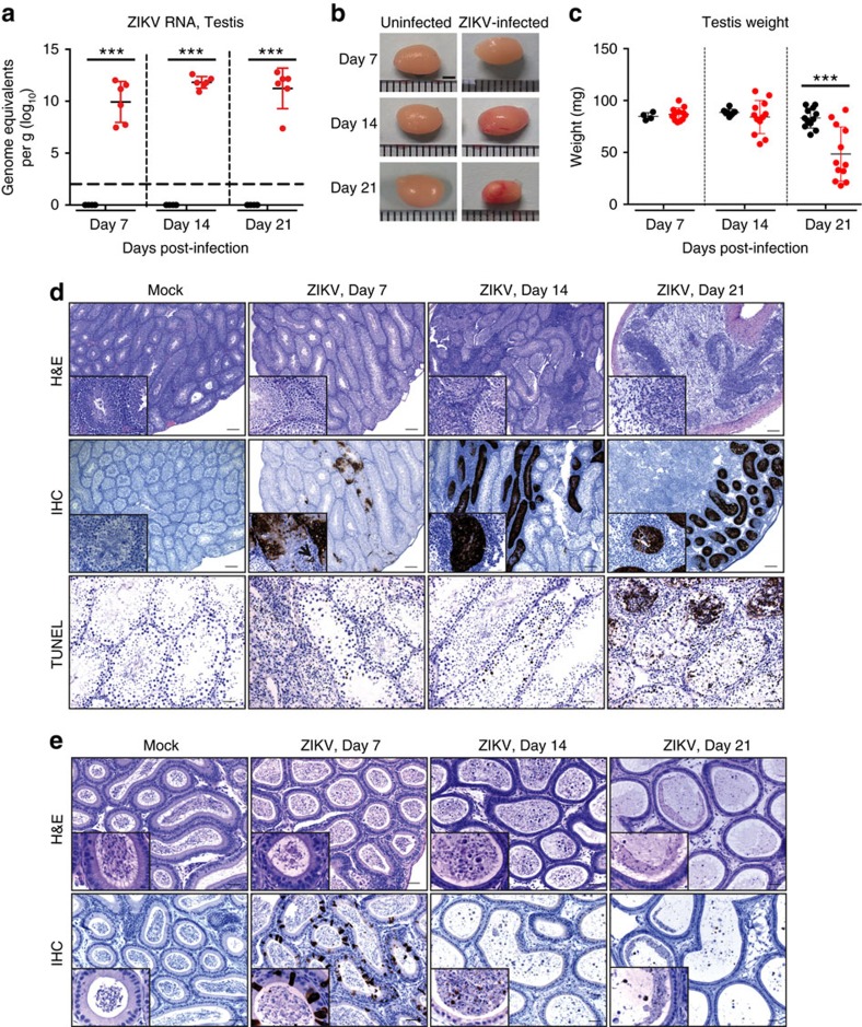
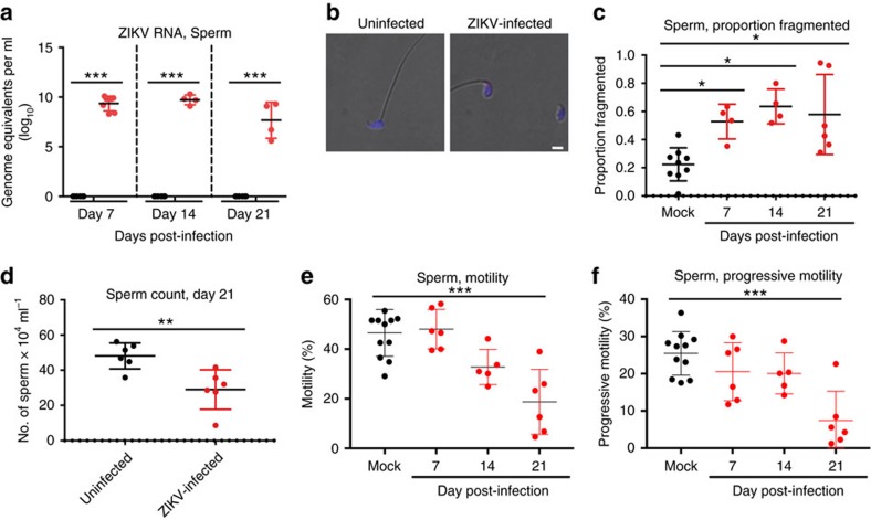
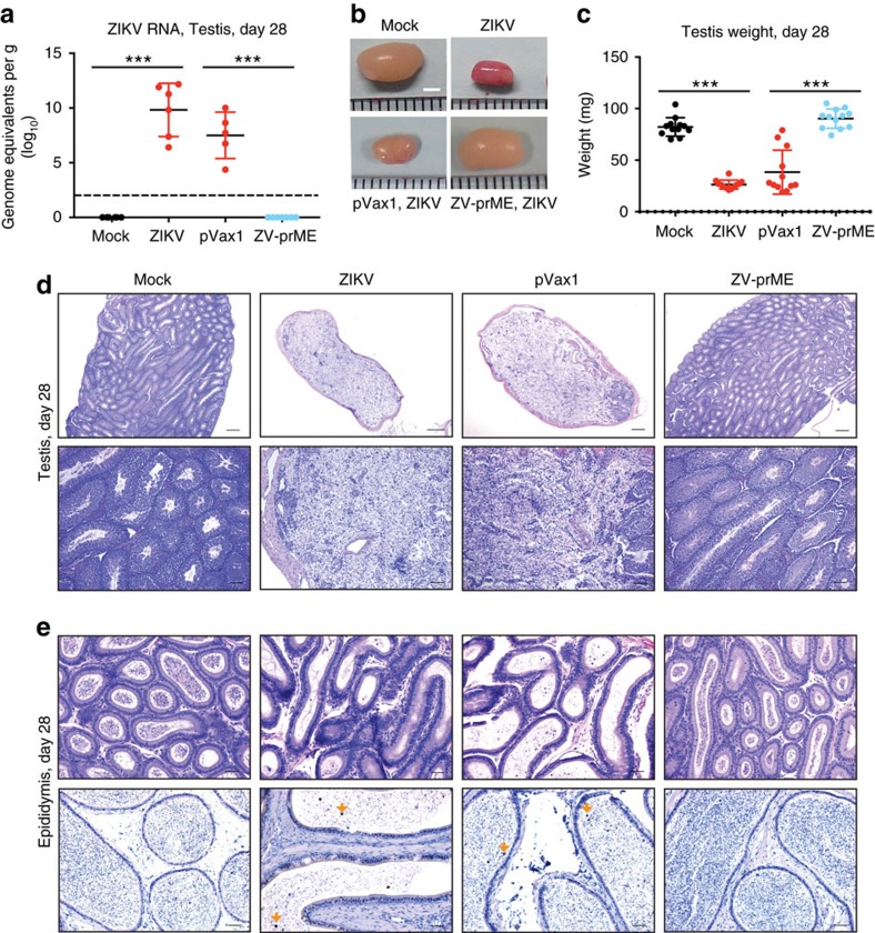
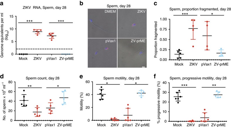
Zika virus (ZIKV) is an emerging pathogen causally associated with serious sequelae in fetuses, inducing fetal microcephaly and other neurodevelopment defects. ZIKV is primarily transmitted by mosquitoes, but can persist in human semen and sperm, and sexual transmission has been documented. Moreover, exposure of type-I interferon knockout mice to ZIKV results in severe damage to the testes, epididymis and sperm. Candidate ZIKV vaccines have shown protective efficacy in preclinical studies carried out in animal models, and several vaccines have entered clinical trials. Here, we report that administration of a synthetic DNA vaccine encoding ZIKV pre-membrane and envelope (prME) completely protects mice against ZIKV-associated damage to the testes and sperm and prevents viral persistence in the testes following challenge with a contemporary strain of ZIKV. These data suggest that DNA vaccination merits further investigation as a potential means to reduce ZIKV persistence in the male reproductive tract.


Conflict of interest statement

D.B.W. has grant funding, participates in industry collaborations, has received speaking honoraria and fees for consulting. This service includes serving on scientific review committees and advisory boards. Remuneration includes direct payments and/or stock or stock options and in the interest of disclosure; therefore, he notes potential conflicts associated with this work with Inovio where he serves on the BOD, Merck, VGXI, OncoSec, Roche, Aldevron and possibly others. The remaining authors declare no conflict of interest.

Figures

Figure 1
Figure 1. ZIKV infection of the testis and epididymis.
Ten- to eleven-week-old male Ifnar1−/− mice were inoculated with 5 × 10⁵ p.f.u of ZIKV strain PRVABC59 by a subcutaneous route (s.c.) into each hind leg (1 × 10⁶ p.f.u. total) and compared to PBS mock-infected mice (n=6). The testes and epididymides were collected at the indicated d.p.i. (a) ZIKV RNA copies in the testis of mice measured by quantitative reverse transcription (qRT)–PCR. Results are expressed as genome equivalents per gram of tissue. The horizontal hatched line indicates the limit of detection (<100 copies per ml). (b) Representative images of testis from mock-infected and ZIKV-infected mice at the indicated d.p.i. (c) Weight of the testis (two per mouse) from mock-infected and ZIKV-infected mice at the indicated d.p.i. (d) Histopathological analysis of the testis (upper panels), immunohistochemical labelling of ZIKV particles in the testis (middle panels), and TUNEL staining of testis (lower panels) at the indicated d.p.i. (e) Histopathological analysis of the epididymis (upper panels) and immunohistochemical labelling of ZIKV particles in the epididymis (lower panels) at the indicated d.p.i. Scale bars, 2 mm in b 200, 200, 50 μm (top, middle and lower panels) in d and 50 μm in e. The data shown are from one experiment that is representative of the same outcome in the two studies performed. Bars indicate mean values and error bars indicate s.d. Statistical differences are given (two-way ANOVA followed by the Bonferroni post-test). ***P<0.001.
Figure 2
Figure 2. ZIKV infection of the sperm.
Ten- to eleven-week-old male Ifnar1−/− mice were inoculated with 5 × 10⁵ p.f.u of ZIKV strain PRVABC59 by a subcutaneous route (s.c.) into each hind leg (1 × 10⁶ p.f.u. total) and compared to age-matched PBS mock-infected mice. Sperm form the caudal epididymis was collected immediately after euthanization. (a) ZIKV RNA copies in the caudal sperm of mice measured by qRT-PCR (n=4 or greater). Results are expressed as genome equivalents per ml. The horizontal hatched line indicates the limit of detection (<100 copies per ml). (b) Immunofluorescence analysis of caudal sperm with anti-ZIKV antibody. Blue indicates DAPI staining and red represents ZIKV. (c) The proportion of fragmented sperm was assessed following staining with Diff-Quick solution. (df) Fertility parameters of caudal sperm were evaluated, including (d) concentration on day 21 after infection and (e) the percentage motility and (f) progressive motility at the indicated times post-infection. Scale bars, 2 μm in b. The data shown are from one experiment that is representative of the same outcome in the two studies performed. Bars indicate mean values and error bars indicate s.d. Statistical differences are given (a, two-way ANOVA followed by the Bonferroni post-test; (c,e,f) one-way ANOVA followed by Dunnett's test; d, Mann–Whitney test). *P<0.05; **P<0.01; ***P<0.001.
Figure 3
Figure 3. Protection from ZIKV-induced damage to the testis and epididymis by DNA immunization.
Five- to six-week-old male Ifnar1−/− mice received two vaccinations by the i.m. route with electroporation-mediated delivery at 2-week intervals with 25 μg of the control vector (pVax1) or ZIKV-prME DNA vaccine (ZV-prME). Mice were challenged 2 weeks after the second immunization with a total of 1 × 10⁶ p.f.u. of ZIKV strain PRVABC59 by a subcutaneous route (s.c.) and compared to age-matched PBS mock-infected (mock) or ZIKV-infected mice (ZIKV). The testes and epididymides were collected at 28 d.p.i. (a) ZIKV RNA copies in the testis of mice measured by qRT-PCR (n=5 or greater). Results are expressed as genome equivalents per gram of tissue. The horizontal hatched line indicates the limit of detection (<100 copies per ml). (b) Representative images of testis. (c) Weight of the testis (2 per mouse). (d) Histopathological analysis of the testis. (e) Immunohistochemical labelling of ZIKV particles in the epididymis. Arrows indicate cells positive for ZIKV antigen. Scale bars, 2 mm in (b), 200 μm in (d, top panel), 50 μm in (d, lower panel), 50 μm in (e, top panel), 10 μm in (e, lower panel). The data shown are from one experiment (n=6) that is representative of the same outcome in the two studies performed. Bars indicate mean values and error bars indicate s.d. Statistical differences are given (two-way ANOVA followed by the Bonferroni post-test). ***P=0.001.
Figure 4
Figure 4. Protection from ZIKV-induced sperm damage by DNA immunization.
Five- to six-week-old male Ifnar1/ mice received two vaccinations by the i.m. route with electroporation-mediated delivery at 2-week intervals with 25 μg of the control vector (pVax1) or ZIKV-prME DNA vaccine (ZV-prME). Mice were challenged 2 weeks after the second immunization with a total of 1 × 10⁶ p.f.u. of ZIKV strain PRVABC59 by a subcutaneous route (s.c.) and compared to age-matched PBS mock-infected or ZIKV-infected mice. Sperm form the caudal epididymis was collected immediately after euthanization. (a) ZIKV RNA copies in the caudal sperm of mice measured by qRT-PCR (n=5). Results are expressed as genome equivalents per ml. The horizontal hatched line indicates the limit of detection (<100 copies per ml). (b) Immunofluorescence analysis of caudal sperm with anti-ZIKV antibody. Blue indicates DAPI staining and red represents ZIKV. (c) The proportion of fragmented sperm was assessed following staining with Diff-Quick solution (n=4). (df) Fertility parameters of caudal sperm were assessed (n=5), including concentration (d), percentage motility (e) and progressive motility (f). Scale bars, 2 mm in b. The data shown are from one experiment (n=6) that is representative of the same outcome in the two studies performed. Bars indicate mean values and error bars indicate s.d. Statistical differences are given (a,cf, one-way ANOVA followed by Dunnett's test. *P<0.05; **P<0.01; ***P<0.001.

References

    1. Coyne C. B. & Lazear H. M. Zika virus—reigniting the TORCH. Nat. Rev. Microbiol. 14, 707–715 (2016). - PubMed
    1. Mlakar J. et al. Zika virus associated with microcephaly. N. Engl. J. Med. 374, 951–958 (2016). - PubMed
    1. Cugola F. R. et al. The Brazilian Zika virus strain causes birth defects in experimental models. Nature 534, 267–271 (2016). - PMC - PubMed
    1. Garcez P. P. et al. Zika virus impairs growth in human neurospheres and brain organoids. Science 352, 816–818 (2016). - PubMed
    1. Miner J. J. et al. Zika virus infection during pregnancy in mice causes placental damage and fetal demise. Cell 165, 1081–1091 (2016). - PMC - PubMed

Publication types